Article Text

Abstract
Objective To assess feasibility and compare the effects of 96-hour shipment of Descemet membrane endothelial keratoplasty (DMEK) grafts as a scroll or a tri-fold on cell viability.
Methods and analysis DMEK grafts were prepared at the Rocky Mountain Lions Eye Bank. Twenty pre-stripped DMEK grafts, paired from 10 donors, were either tri-folded in an endothelium-in configuration using microforceps and loaded into a plastic Treyetech cartridge, or suctioned in a scrolled endothelium-out configuration into a modified Jones Tube. Grafts were shipped via FedEx to a secondary location and back for 48 hours each way, resulting in a total shipping time of 96 hours. After shipping, grafts were removed from inserters onto glass slides and unfolded using viscoelastic with endothelium facing upwards. Calcein-AM stained grafts were imaged with a fluorescent microscope and endothelial cell loss (ECL) was measured using trainable segmentation in Fiji by a masked grader.
Results A total of 20 grafts were shipped for 96 hours, split between preloaded tri-folded (n=10) and preloaded scrolled (n=10) tissues. No significant difference in ECL was observed across groups after prolonged shipping (14.8% vs 13.7% ECL respectively, p=0.68).
Conclusion For preloaded DMEK after 96 hours, both scrolled and tri-folded tissue demonstrated clinically acceptable levels of ECL. The data suggest a wider window of time for endothelial cell viability and is promising for the prospect of international shipment of preloaded grafts.
- eye (tissue) banking
- cornea
- DMEK
- preloaded
- trifolded
- descemet membrane endothelial keratoplasty
Data availability statement
All data relevant to the study are included in the article.
This is an open access article distributed in accordance with the Creative Commons Attribution Non Commercial (CC BY-NC 4.0) license, which permits others to distribute, remix, adapt, build upon this work non-commercially, and license their derivative works on different terms, provided the original work is properly cited, appropriate credit is given, any changes made indicated, and the use is non-commercial. See: http://creativecommons.org/licenses/by-nc/4.0/.
Statistics from Altmetric.com
Key messages
What is already known about this subject?
Scroll-based, preloaded Descemet membrane endothelial keratoplasty (DMEK) can be safely stored in place for 5 days or shipped over a 3-day period; however, data is still needed regarding endothelial cell viability for preloaded DMEK grafts shipped beyond 72 hours.
What are the new findings?
This study finds that both tri-folded and scroll-based preloaded DMEK grafts can be shipped over a 96-hour period with acceptable and comparable rates of endothelial cell loss.
How might these results change the focus of research or clinical practice?
This study’s data and results support the possibility of international shipping of preloaded DMEK grafts over longer-time intervals.
Introduction
Relative to previous variations of cornea transplantation, such as Descemet stripping automated endothelial keratoplasty (DSAEK), Descemet membrane endothelial keratoplasty (DMEK) optimises visual outcomes.1 Preloading tri-folded tissue has been proven to successfully facilitate the DMEK procedure by minimising surgical time and costs while retaining clinically acceptable levels of endothelial cell loss (ECL).2 These advancements have shifted graft preparation out of the operating room and into the eye bank.
Preliminary studies have previously tested the effect of shipping on corneal graft health after 48–72 hours.3 For DSAEK, precut grafts have been shown to maintain clinically acceptable ECL after international shipment (mean shipment of 62.3 hours). After 48 hours of shipping, tri-folded DMEK grafts demonstrated a comparable ECL to scrolled DMEK grafts, with a marginally significant trend demonstrating 4.3% less ECL in tri-folded as compared with scrolled grafts.4 5 However, there has been limited investigation into the effect of extended shipping on DMEK grafts with a comparison between these two methods.6
In 2019, over 28 000 corneas were exported internationally from the USA for keratoplasty.7 The ease of said international transplants would be greatly improved if grafts could be preloaded and were to remain viable during prolonged shipping. Therefore, this study sought to determine whether graft viability is maintained for preloaded tri-folded versus preloaded scrolled tissue intended for DMEK surgery during longer shipments. Over 96 hours, transport conditions were simulated by shipping packaged grafts to a secondary location and back.
Materials and methods
Graft procurement
Corneas were recovered by the Rocky Mountain Lions Eye Bank (RMLEB) and were used if they were found ineligible for clinical transplant use and consented for use in research by next of kin. Corneas with scars, wounds or anterior stromal infiltrates were excluded from the study. A certified eye bank technician verified, via slit lamp evaluation, that corneas used had no worse than mild localised ECL or damage, and the endothelium of each cornea was verified to be reflective and have visible cell borders. Endothelial cell density was measured using the Konan CellChek D specular microscope for each graft at this initial stage, prior to testing.
Immediately before DMEK prep, each cornea pair was removed from its storage medium (Optisol-GS; Bausch & Lomb, St Louis, Missouri, USA or Life4C; Numedis, Isanti, Minnesota, USA) and stained for 30 s–1 min with a 0.06% solution of trypan blue (VisionBlue; Dutch Ophthalmic, Exeter, New H) to identify any further damage that would prevent use for the study.
Experimental groups
A total of 20 corneas were divided into two experimental groups with 10 corneas each. The grafts were peeled according to standard eye bank protocol and then loaded in a scroll into either a modified Jones Tube for DMEK (80000-DMEK; Gunther Weiss Scientific Glassblowing, Portland, Oregon, USA) or tri-folded in a plastic Treyetech cartridge (Xcentric Mold & Engineering, Shelby Township, Michigan, USA).8 9 The grafts were then shipped for 96 hours before unloading and staining (n=10, each device).
In order for each pair of corneas to serve as its own internal control, corneas from the same donor were split between the two cartridge types (eg, the left cornea was used with the Jones Tube, and the right cornea from the same donor was used with the Treyetech cartridge). In total, all 10 grafts in each cartridge group originated from a paired donor.
Graft peeling
DMEK grafts were prepared by one eye bank technician according to standard RMLEB procedures outlined in Barnes et al.4 After using a 9.5 mm diameter guarded corneal punch (Moria SA, Antony, France), to punch the Descemet membrane (DM), the resulting wound was visualised using a short (30 s–1 min) 0.06% trypan blue solution staining. Working under a pool of corneal storage medium, the outer piece of DM was removed, leaving the central 9.5 mm DM disk. A Sinskey hook was used to separate the peripheral edge of the 9.5 mm DM disk from the stroma for approximately 350 degrees, leaving a small peripheral attachment. This peripheral attachment was oriented to 6 o’clock, and the 12 o’clock edge of the DM disk was grasped with the ‘heel’ portion of curved tying forceps and carefully peeled from the stroma by pulling toward the peripheral attachment until approximately 90% of the DM disk was separated. The peeled DM was carefully laid down in the storage medium. Using fluid movement and capillary action, the DM disk was returned to its original position on the stroma in the storage medium by drawing the fluid toward one side using a sponge. Excess storage medium was removed to ensure a flat, unscrolled position. The 9.5 mm disk was then cut to 8.0 mm diameter surgical graft size using a standard corneal punch (Moria SA, Antony, France), followed by 2–4 min of 0.06% trypan blue solution application ensuring stromal side exposure.
Graft loading and shipping
Method 1: modified Jones Tube (scroll method)
The remaining anterior cornea was used to carry the separated 8.0 mm DMEK graft and was transferred to a glass petri dish containing storage medium, either Optisol-GS or Life4C. The storage medium preferred by the eye bank was transitioning from Optisol-GS to Life4C through the study. However, all paired grafts were stored in identical solution so that no difference existed in storage medium between groups, minimising impact on our analysis. The graft was encouraged to scroll and float. At this point, the anterior cornea was removed without direct instrument to graft contact. Once the graft had sufficiently scrolled, it was aspirated into a modified Jones Tube filled with fresh, cold storage medium. This configuration is shown in figure 1A.
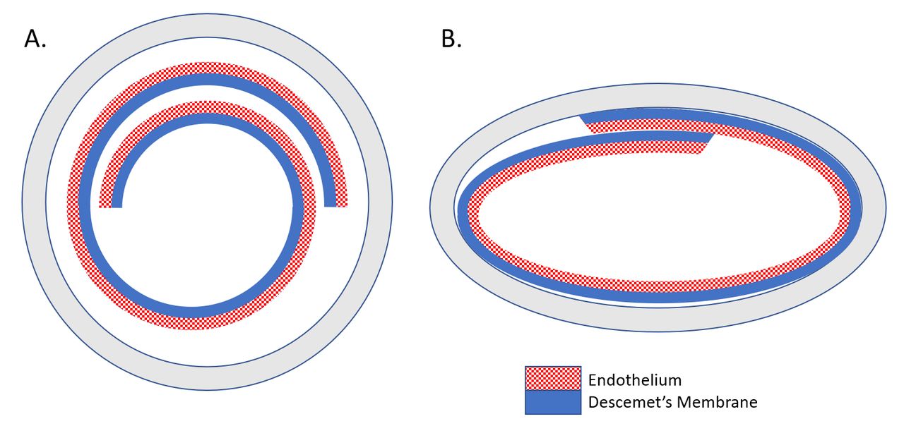
Graft configuration in Jones Tube and Treyetech cartridge. Endothelium is represented by the chequered pattern and stroma is represented by the solid pattern. (A) Grafts in the Jones Tube are in a scrolled endothelium-out configuration. (B) Grafts in the Treyetech cartridge are in a tri-folded endothelium-in configuration.
The 10 grafts were shipped from RMLEB using FedEx under routine transplant tissue shipping cool conditions, packaged with water ice. After arriving at their destination on the East Coast of the USA at the 48-hour mark, fresh ice was added to the packaging and the grafts were shipped back to their original location for an additional 48 hours. After shipping, the grafts were ejected onto a bed of Calcein-AM (Invitrogen, Thermo Fisher Scientific, Waltham, Massachusetts, USA) and Amvisc Plus (Bausch+Lomb, Rochester, New York, USA) and unfolded without direct contact via the use of viscoelastic.
Method 2: tri-fold method
The DMEK graft peripheral attachment was separated with a Sinskey hook by sliding gently under the membrane, and two or three drops of 0.06% trypan blue solution were added. The graft edges were freed from the punch wound using a Sinskey hook to slightly lift the edges, allowing trypan to flow into the interface between DM and the stroma. After approximately 2 min, the graft was carefully tri-folded using forceps with the endothelium on the inside of the fold. The tri-folded graft was then transferred into the Treyetech cartridge using microforceps and secured stably into the inserter with fresh, cold storage medium, either Optisol-GS or Life4C. This configuration is shown in figure 1B. Paired grafts were stored in identical solution so that no difference existed in storage medium between groups, minimising impact to analysis.
The 10 grafts were shipped from RMLEB using FedEx under routine transplant tissue shipping cool conditions, packaged with water ice. After arriving at their destination on the East Coast of the USA at the 48-hour mark, fresh ice was added to the packaging and the grafts were shipped back to their original location for an additional 48 hours. After shipping, the graft was pulled from the front cartridge using microforceps onto a bed of Calcein-AM and Amvisc Plus and unfolded without direct contact via the use of viscoelastic.
Staining and imaging
Fluorescence staining was performed with a 1.7 µM solution of Calcein-AM in balanced salt solution (BSS) by a single eye bank technician to facilitate checking for cell viability after the grafts were removed following shipping. Amvisc Plus viscoelastic was used to unscroll the graft without direct instrument manipulation for imaging. After 20–40 min of Calcein-AM exposure, several images of the graft were captured using a Leica DM IL inverted fluorescence microscope with an attached AmScope model MT5000(IFR) CCD.10 GIMP was used to stitch images from each graft into one whole image per graft.11
These images were analysed by a masked grader who was uninvolved in DMEK graft preparations using Fiji ImageJ12 with trainable segmentation. The segmentation was trained to return the total ECL of each imaged graft calculated as the percentage of non-viable graft area.
Statistical analysis
To test for a difference in mean per cent ECL across DMEK graft configurations, a paired t-test was conducted between the scroll and tri-folded group. The effects of donor characteristics on ECL were minimised by using paired grafts from the same donor for the two experimental groups. The difference in per cent ECL of the grafts from each unique donor was used to determine statistical significance.
Patient and public involvement
There was no patient or public involvement in the design or execution of this study.
Results
A total of 20 grafts were used in this study. The age range of donors was 44–74 years (mean, 63.3 years, median 67.5 years), the death-to-preservation time ranged from 03:26 to 16:10 hours (mean 10:38 hours, median, 10:34 hours) and the death-to-preparation time ranged from 2 to 17 days (mean 8.4 days, median 8.5 days).
These grafts were divided into two groups, scrolled in the Jones Tube (n=10) or tri-folded in the Treyetech cartridge (n=10). Prior to preloading and shipping for 96 hours, there was no significant difference in mean endothelial cell density between tissues that were scrolled or tri-folded (2626 vs 2575 cells/mm2, p=0.82).
After 96 hours, no grafts in either group had been ejected from their cartridges. The mean total ECL of all grafts following transport was 14.2%. When comparing across groups, the rates of total ECL after transport among grafts shipped in the Jones Tube was found to be 13.7% (95% CI, 10.9% to 16.4%), while the mean total ECL of the tri-folded grafts shipped in the Treyetech cartridge was 14.8% (95% CI, 11.1% to 18.5%). As the grafts in each group originated from a paired donor and were shipped under identical conditions (eg, storage medium), a paired t-test between groups of the mean ECL after 96-hour shipping was performed. The paired t-test revealed no significant difference in the mean total ECL across the two experimental groups (p=0.68). The total ECL from each graft appears in table 1.
Endothelial cell viability in 20 paired, preloaded donor grafts for Descemet membrane endothelial keratoplasty (DMEK) shipped for 96 hours. Both tri-folded and scrolled DMEK grafts demonstrated comparable and acceptable rates of endothelial cell viability
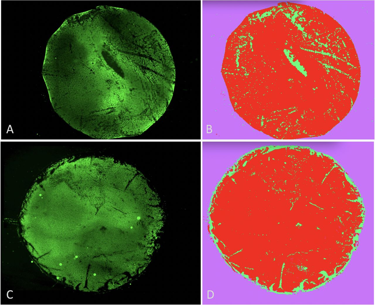
Images were then analysed for qualitative differences in ECL. Among DMEK grafts shipped as a scroll, several grafts demonstrated central linear regions of ECL, consistent with glass-graft contact. In contrast, ECL in tri-folded grafts was frequently identified at the peripheral edge, where grafts are handled during the folding and pulling process. These patterns are depicted in figure 2.

Representative images of stained and segmented grafts after shipping and removal from cartridges. (A) Fluorescent image of donor 2’s graft after immediate removal from the modified Jones Tube and staining. (B) Segmented image of donor 2’s graft, showing cells classified as viable in red. Measured endothelial cell loss (ECL) is 11.4%. (C) Fluorescent image of donor 7’s graft after immediate removal from the Treyetech cartridge and staining. (D) Segmented image of donor 7’s graft, showing cells classified as viable in red. Measured ECL is 10.8%.
Discussion
This study demonstrates that preloaded DMEK grafts can be shipped for at least 96 hours, stored in Optisol or Life4C and on ice, with clinically acceptable levels of ECL. A comparison between configurations (scrolled vs tri-folded) of paired preloaded grafts in identical media revealed a non-significant difference in ECL over a 96-hour shipping period.
While many preloaded tissues are delivered within 24–48 hours of preparation, in vitro studies of ECL with 72 hours of storage as a preloaded scroll have suggested that tissue may be able to be sustained for a longer period of time, with rates of ECL ranging from 15% to 22%.13 14 In this study, shipping time was extended beyond such studies by an additional 24 hours without evidence of additional endothelial compromise, as ECL rates measured across groups averaged under 15%.
In comparing ECL across research groups or eye banks, variability may exist in preparation methods or tissue handling, resulting in slight differences in rates of ECL using the same technology. Barnes et al previously reported ECL using similar methodology in 40 grafts after 48 hours. In this study, an ECL of 11.6% in the tri-folded group and 16.6% in the scrolled group was reported. This suggests that the 96-hour interval does not appear to contribute substantial damage to the grafts beyond what has been demonstrated at 48 hours.6 15 16
The longer time interval allowed for shipping in this study may build opportunities for clinical collaboration across large geographical distances. The data supports the proposal that grafts could be prepared, either both in scrolled or tri-folded configurations, before being shipped or carried by air travel across countries, supporting surgeries in more isolated regions. Additionally, surgeons may benefit from the decreased scrolling tendency of preloaded tri-folded tissue,17 but future studies would need to assess the extent to which this is a time-dependent process. Preloaded systems will facilitate DMEK procedures and decrease the burden on surgeons to prepare their own tissue.
A limitation of this study, shared with most in vitro DMEK experiments, is that the limited number of grafts may result in low statistical power to discern differences between groups. However, we can increase the quality of the comparison by ensuring that the grafts are comparable between the two arms of the study. In this assessment, two approaches were used to optimise such a comparison. First, we used paired grafts from the same donor, to reduce the risk of confounding variables. Second, we performed measurements of endothelial cell density prior to the experiment in all grafts to ensure that the two grafts correlated in their overall health.
In summary, the results of this study confirm that DMEK grafts can be safely preloaded and shipped in cartridges over 96 hours in both scrolled and tri-folded configurations.
Data availability statement
All data relevant to the study are included in the article.
Ethics statements
Ethics approval
Donor tissue used in this study was recovered by the Rocky Mountain Lions Eye Bank and was consented for use in research by next of kin.
Footnotes
Contributors All persons who meet authorship criteria as recommended by ICMJE are listed as authors. The contributions of the authors satisfying all four given conditions are listed specifically below. CONDITION 1: Substantial contributions to the conception or design of the work: CC, JL, EC, KAB, AC, AOE or Substantial contributions to the acquisition, analysis or interpretation of data: CC, SJS, JL, ST, SB, BGW, DSL, EC, KAB, AC, AOE. CONDITION 2: Drafting the work: CC, SJS, SB, BGW, DSL, EC, KAB, AC, AOE or Revising it critically for important intellectual content: JL, ST, AOE. CONDITION 3: Final approval of the version published: CC, SJS, JL, ST, SB, BGW, DSL, EC, KAB, AC, AO E. CONDITION 4: Agreement to be accountable for all aspects of the work in ensuring that questions related to the accuracy or integrity of any part of the work are appropriately investigated and resolved: CC, SJS, JL, ST, SB, BGW, DSL, EC, KAB, AC, AOE.
Funding This work was supported by Research to Prevent Blindness (Special Scholar Award) and the National Institute of Biomedical Imaging and Bioengineering (Design by Biomedical Undergraduate Teams).
Competing interests CC, EC, KAB and AOE have ownership interest in Treyetech.
Patient and public involvement Patients and/or the public were not involved in the design, or conduct, or reporting, or dissemination plans of this research.
Provenance and peer review Not commissioned; externally peer reviewed.