Article Text

Statistics from Altmetric.com
Basal cell carcinoma, the most common periocular malignancy, most commonly affects the lower eyelid and medial canthus and may involve the lower canaliculus.1 Management poses several surgical challenges due to the vicinity’s intricate and complex anatomy.2 Sacrifice of a part of the proximal lacrimal drainage system is not uncommon when the medial portions of the eyelids are involved. Based on a retrospective review of 22 eyes, Chiu et al posit that marsupialisation of the remnant canaliculus after losing part of the proximal lower canaliculus will help prevent postoperative epiphora.3 We have all long faced this problem with what to do with the injured canaliculus and, to date, none of us has the answer. So it behoves us to consider this study and review its conclusions critically.
Tear physiology and drainage are a complex interplay of several factors like the punctal size, position and patency, a normal caruncle and plica, canalicular patency, the blink, the lacrimal pump mechanisms and sinonasal factors.4 Also, the eyelid architecture, by definition, will be different after tumour resection and reconstruction. A variable combination of these factors can be affected adversely following a medial canthal tumour excision. Besides, several new factors like poor quality of ocular surface lubrication, subtle eyelid malposition following reconstruction, and tear film instability can contribute to epiphora. Hence, it is essential to recognise that epiphora in such patients is likely to be multifactorial.
The obvious first question is whether anything needs to be done at all, as there has long been an argument in the oculoplastic world that loss or injury to one canaliculus will not necessarily lead to epiphora. An oft-repeated saw in ophthalmology is that of Abraham Werb from 1987, ‘The conservation of the inferior canaliculus is for tear drainage and the superior canaliculus for the ophthalmologist’,5 illustrating the long-held belief of the lower canaliculus being the main output of tears. Furthermore, although the majority of basal cell carcinoma recurrences occur within 3 years following treatment, 18% reappear between the fifth and tenth year. So conventional wisdom has dictated that no primary lacrimal system reconstruction be done, although there has been no established guideline based on studies.
Carter et al6 found the lower punctum diameter to be 0.321 mm2 and the upper diameter 0.264 mm2 with no significant differences in the sizes between laterality or genders. Based on this, they proposed that the larger size and diameter of the lower punctum would mean that drainage of tears through the lower lacrimal system would be higher than through the upper one. Bothra et al7 recently confirmed this difference in the size of the upper and lower puncta using Dournier-domain optical coherence tomography from the first to the eighth decade of life, but they felt they could not comment on the relative flow of tears from the upper and lower puncta and canaliculi based on their study. In a study of student nurses, Jones and Wobig showed that transit time of tears through the superior and inferior canaliculi is nearly equal.8 Indeed, dacryoscintigraphy has shown no statistical difference in tear flow between the upper and lower canaliculi.9 10
If one canaliculus is obstructed and the eyelids are in good position, it has been shown that tears will flow effectively through the intact punctum and canaliculus without symptoms of epiphora.11 There is also evidence that if one canaliculus is blocked the other uninvolved canaliculus may increase the drainage of tears flowing through it.12 Furthermore, it is also known that there is variability of tear flow from patient to patient. Some patients may drain more tears from the upper or the lower lacrimal systems and it is unpredictable as to whether any individual patient will have tearing if a lacerated canaliculus is left unrepaired.13 Ortiz and Kraushar found that 75% of patients in whom the lower canaliculus repair failed did not develop a watery eye.14 Smit and Mourits found no epiphora in 16 patients with a monocanalicular injury where no surgical reconstruction was performed.15
In summary, although tear outflow is usually similar between the upper and lower canalicular systems, canalicular dominance can vary between individuals and between eyes. Less than 10% of monocanalicular patients experience epiphora in basal tear settings.16–18 It has been proposed that an autoregulatory mechanism compensates for dynamic changes in the tear system.19 20 Yen et al21 found that punctal occlusion affects the interaction between the ocular surface and the lacrimal gland and decreases tear production in normal subjects. They postulated that, if the effect of mono-occlusion of the canaliculus exceeds the compensation of the ipsilateral, non-occluded canaliculus, the tear system would respond by decreasing tear secretion to maintain the appropriate tear quantity. Linberg and Moore16 used temporary punctal plugs to create monocanalicular obstructions. They found that a single upper or lower canaliculus was inadequate to drain reflex tear secretion completely without symptoms in 50% of patients tested.
Just as it is now accepted that all canalicular lacerations should be repaired whenever possible without risking the integrity of the uninjured canaliculus, we should make every effort to improve tear egress in cases after tumour resection. It is, of course, vital that the intact opposite canaliculus and punctum should not be injured during any reconstructive procedure. This is the reason why a primary bicanalicular intubation at the time of the repair may be unwise as it carries the risk of injuring the intact punctum or canaliculus. However, one could argue that the risk would presumably be low enough to consider this option as it would at least keep the injured canaliculus ‘open’ against the inevitable scarring that would occur around the site of eyelid reconstruction. How low a risk is acceptable? We don’t know. If a patient ends up with epiphora with one functioning canaliculus, it has been shown that a dacryocystorhinostomy will help many of these patients prior to resorting to the insertion of a Jones tube.22
Next, we must ask about the procedure of marsupialisation and its effect on the function of the canaliculus. Older, suggested opening the superior part of the remaining canaliculus by 3–4 mm and suturing the edges of the opened mucosal lining to the surrounding tissues.23 In this paper, the authors open ‘the entire remnant canaliculus’. They do not state if they used sutures to keep the marsupialised canaliculus ‘open’. Marsupialising a canaliculus is somewhat akin to the slit canaliculi we see in clinical practice, except that in a slit canaliculus, there is generally no total loss of any of the punctum or the canaliculus. Most of the surrounding innervation, fascia and musculature would be intact. It has been shown that a slit canaliculus does not cause a functional problem in most patients.24
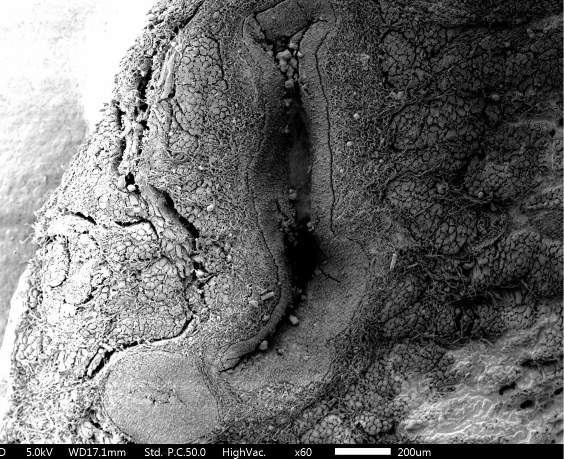
How may marsupialisation affect the function of the canaliculus? With each blink, the pretarsal orbicularis oculi muscle compresses the ampullae, shortens and compresses the horizontal canaliculi, and closes and moves the puncta medially, resisting reflux. Simultaneously, contraction of the lacrimal part of the orbicularis oculi creates a positive pressure that forces tears down the nasolacrimal duct and into the nose, mediated by helically arranged connective tissue fibres around the lacrimal sac. When the eyes are opened, the canaliculi and sac expand, creating negative pressure that draws tears from the canaliculi into the sac. Recent electron microscopy and three-dimensional histology have demonstrated that at its insertion, the Horner-Duverney’s muscle continues to encircle the canaliculi in a scissoring pattern, which gets exaggerated around the vertical canaliculus and becomes denser and more parallel as it continues around the horizontal canaliculus (figure 1).25 Loss of the proximal part of the lower canaliculus results in compromise of the physiological functions of this part of the lacrimal system. The already compromised driving force that creates a negative pressure within the canaliculi will probably be further compromised with a distal marsupialisation. Besides, there is a risk of further traumatising the heavy neural innervation in the pericanalicular musculature with subsequent functional implications. Marsupialisation will inevitably cause some change in the capillary action in a tube that occurs more strongly in narrow bore tubes than in wider ones. It can be postulated that some of the capillarity would be affected by changing the bore structure of the canaliculus into an open receptacle. The Venturi tube effect increases the movement of tears as the canaliculus narrows with eyelid blinks. This presumably would be weakened or lost with marsupialisation. One assumes that marsupialisation is simply avoiding constriction that would occur with scarring, were nothing to be done to the cut canaliculus and therefore acting simply as a lacus for the tears and relying on the rest of the physiological processes to propel the tears onward. Can one determine if there is, indeed, better flow of tears once a traumatised canaliculus has been marsupialised? Unfortunately, neither dacryoscintigraphy nor dacryocystography would allow one to determine if there is improved flow through the marsupialised canaliculus. Lacrimal pump dysfunction, canalicular fibrosis, and fistula formation are documented complications of canaliculotomy procedures.26 27 With these considerations in mind, it is reasonable to say that marsupialisation of the remnant canaliculus is a relatively simple procedure that can be performed at the time of reconstruction after tumour resection. It also avoids manipulation of the intact punctum and canaliculus, which is important.
Scanning electron microscopy image of the lacrimal canaliculus showing its intricate relation with Horner-Duverney muscle. The physiological implications of the dense autonomic and sensory innervation seen still remain to be deciphered.
This stuy by Chiu et al does not grade tearing nor does it assess reflex tearing. It is because the loss of one canaliculus causes most patients not to experience epiphora that a robust control group would have made the results more meaningful.
It is assumed in the paper that all repairs would be equally good in terms of the apposition of the reconstructed eyelid to the globe and the excursion and meeting of the upper eyelid to the lower eyelid in a blink, which would allow the movement of tears with each blink through the uninjured punctum and canaliculus. Of course, this can never be the case, as it is the nature of loss of tissue from tumours that involve part of the lower lacrimal system and eyelid that there will be some repairs that work better than others. And this will also have an effect on the possibility of epiphora. There is also the inevilability that all of us who work in teaching institutions face: some patients will have been operated on by residents or fellows. Even with appropriate supervision, this does not fall under the purview of ‘single surgeon surgery’ results, which obviously has advantages.
In this series, the mean age was 77.6 years. A significant proportion of these patients will have dry eyes, some degree of delay in lacrimal outflow, or even asymptomatic obstruction. Documenting the presence or absence of dry eyes and patency of the lacrimal system prior to any surgery would make such a study more robust.
In a review of the literature, Chiu et al found the rate of epiphora where no reconstruction of the injured canaliculus was performed to be 12.5% (of 97 eyes). The authors found epiphora in 9.1% of their 22 patients who underwent marsupialisation.3 One can certainly not claim that marsupialisation of the damaged canaliculus definitively gives superior results in terms of epiphora based on these results. However, and not withstanding the discussion about our more recent understanding of canalicular anatomy and physiology and the possible disturbance of the physiological forces that propel tears along a canaliculus, it may be stated that marsupialisation, as far as we know, subscribes to the Hippocratic oath, primum non nocere, and, may actually be helpful. All things considered, based at least on the study as presented, it is soft evidence at best. A perfect study this may not be, but this discussion is not to impugn this perfectly sensible effort. As we seek to improve our results and our patients’ lives, such studies can lead to more robust controlled studies and prove (or disprove) the soft evidence presented. That is all we have to say.
Ethics statements
Patient consent for publication
References
Footnotes
Contributors MJA: produced the initial outline of an editorial response to the article by providing histopathological original work in the form of a scanning electron microscope and wrote the description of the anatomy of the punctum and canaliculus based on his original work. GER: provided an analysis of the results of his experience in treating tumours which involve the canaliculus over 30 years and distilled this into an informed opinion about the use of marsupialisation in the presence of loss of part of a canaliculus. RM: did the original research on tumours of the eyelids in Australia and we based some of our observations on this original work of his. BCKP collected all the opinions, performed the research of the subject and wrote the final version and added his opinions to the editorial.
Funding MJA receives royalties from Springer for the textbook 'Principles and Practice of Lacrimal Surgery’ and Atlas' 'Atlas of Lacrimal Drainage Disorders' and the ‘Video Atlas of Lacrimal Drainage Surgery’. BCKP is supported in part by an Unrestricted Grant from Research to Prevent Blindness, New York, NY, to the Department of Ophthalmology and Visual Sciences, University of Utah. He also receives royalties from Springer for the textbook 'Clinical Ophthalmic Oncology-Orbital Tumours'.
Competing interests None declared.
Provenance and peer review Commissioned; internally peer reviewed.
Linked Articles
- Orbit and Oculoplastics