Article Text

Abstract
Objective To detect the presence of microorganisms in the storage media of human donor corneas using next-generation sequencing method.
Methods Seven samples from organ culture (OC) group (Cornea Max, Eurobio, Les Ulis, France) with one control (sterile media without any cornea) and seven samples from hypothermic storage group (Cornea Cold, Eurobio) with one control were used for this study. The corneas were placed in the respective storage media for 14 days before collecting the samples. Storage media (2 mL) from each sample were collected in RNAase-free tubes and shipped for ribosomal RNA sequencing of 16 S and 18 S. Simultaneously, another 1 mL of media sample was used for conventional diagnostic method (CDM) using Bactec instruments.
Results In both, OC and hypothermic storage and control samples, the most abundant genera were Pseudomonas, Comamonas, Stenotrophomonas, Alcanivorax, Brevundimonas and Nitrobacter. Acidovorax, Acetobacter and Hydrogenophilus were detected mostly in the hypothermic storage group. The most abundant fungal pathogen detected belonged to the genus Malassezia, which was found in both the storage conditions. CDM was negative for microorganisms in all the samples.
Conclusion Metagenomics provides full taxonomic profiling of the detected genomic material of the organisms and thus has the potential to deliver a much wider microbiological diagnostic approach than CDM. The costs and turn-around time need to be reduced, and; the detection of viable organisms would help this technology to be introduced into routine clinical practice.
- cornea
- eye bank
- microbiology
- NGS
- 16S
- 18S
- bacteria
- fungus
- storage
- preservation
- media
This is an open access article distributed in accordance with the Creative Commons Attribution Non Commercial (CC BY-NC 4.0) license, which permits others to distribute, remix, adapt, build upon this work non-commercially, and license their derivative works on different terms, provided the original work is properly cited, appropriate credit is given, any changes made indicated, and the use is non-commercial. See: http://creativecommons.org/licenses/by-nc/4.0/.
Statistics from Altmetric.com
Key messages
What is already known about this subject?
PCR techniques for 16S and 18S, and shotgun sequencing approach has already been described in the earlier reports investigating its use in the field of ocular surface infections and identifying its microbiome. This is the first paper reporting about the use of metagenomics in eye banking field.
What are the new findings?
This study investigates the presence of microorganisms by detecting the genomic material present in the corneal preservation medium. The results suggest that both, 16S and 18S techniques require less amount of samples and are efficient in identifying the genomic material in the preservation media with better taxonomical profiling.
How might these results change the focus of research or clinical practice?
This approach could be useful as it requires less amount of starting sample, which has always been a routine issue, and provides complete taxonomical profile of an organism compared with the ongoing conventional diagnostic methods, that only reports the presence or absence of an organism. This study will be useful in the research and clinical practice in terms of its high specificity, complete profiling and hypothesis-free approach. However, the costs, turn-around time and downstream analysis of the data still needs to be improved for the technology to be used routinely.
Introduction
Infections of the eye such as endophthalmitis may occur following a corneal transplant. The incidence of endophthalmitis over a 7-year period in the UK following penetrating keratoplasty was 0.67%.1 It can be challenging to identify and distinguish the source of the infection, which includes endogenous source such as the host and a variety of exogenous sources such as the donor cornea. It is well known that donor corneas may be the source of contamination as they contain viable cells and as such cannot undergo typical sterilisation processes. Risk factors for the development of an infection following a corneal transplant include immunosuppressive treatment following surgery and cornea’s avascular state.2 It has been suggested that discontinuation of the topical antimicrobials with concomitant use of steroids may allow growth of sequestered microorganisms. Transmission of the herpes simplex virus type 1 from the donor cornea has shown to increase the risk of rejection.3 Fungal infections have also been reported in the preservation media affecting graft survival after transplantation.4 5 The specific diagnosis of infection remains a challenge as it still relies on conventional microbial culture techniques for the identification of the suspected pathogen. Most of the environmental microorganisms that are difficult to culture using conventional techniques can be detected using molecular methods. Such techniques use a hypothesis-free short-read approach that is suited for taxonomic and functional profiling applying the high-throughput DNA sequencing (DNA-seq) techniques. This helps to determine microbes with low sample volume increasing the diagnostic yield. PCR analysis has already been applied to identify pathogenic agents in ocular tissues, including the aqueous humour and vitreous, and has been used for the diagnosis of infections that would have been otherwise difficult to identify.6 RNA sequencing can also be performed to detect fungus, parasites and viruses7; however, this technique has limitations, as it requires proper specimen handling. For pathogens with DNA genomes, metagenomic DNA-seq can circumvent this challenge, as DNA is more stable at ambient temperatures. In this study, we investigate whether there are microorganisms present in the storage media that are undetected using the conventional microbiological assays.
Materials and methods
Ethical statement
Human donor corneas were obtained by the Veneto Eye Bank Foundation, Venice, Italy, with written consent from the donor’s next-of-kin to be used for research purposes. The study followed the 2013 Tenets of Declaration of Helsinki. The tissues were used under the laws of Centro Nazionale Trapianti, Rome, Italy. The corneas were suitable for research and unsuitable for transplantation due to low endothelial cell counts (<2200 cells/mm2). No other complications or indications were recorded in the donor corneas such as diabetes, HIV or hepatitis B virus.
Corneal preservation
Human donor corneas (n=7) were excised and preserved in Cornea Max (Eurobio, Les Ulis, France) for 14 days at 31°C, that is, the protocol currently used for organ culture (OC). Other (n=7) samples were preserved in Cornea Cold (Eurobio) for 14 days at 4°C, as current hypothermic protocol. Both the media are commercially available and contain penicillin and streptomycin as antibacterial agents and amphotericin B as an antifungal agent. The control samples (n=1) from OC and hypothermic media, without human donor corneal tissues, were used separately as controls.
Sample collection from corneal preservation media
Two millilitres of the storage media from each sample was extracted and preserved in 2 mL sterile Eppendorf tubes (Eppendorf Biopur safe-lock microtubes, Sigma-Aldrich, Italy) and shipped to IMGM laboratories, Germany, at room temperature (OC media) and in dry ice (hypothermic media), for metagenomic analysis, respectively. Microbiological analyses were also carried out in-house on the same samples using a Bactec Instrument (Becton Dickinson, Franklin Lakes, NJ, USA), which is a colorimetric assay, in order to compare the difference between metagenomic analysis and conventional eye banking microbiological tests.
DNA isolation
Genomic DNA (gDNA) was isolated from both the preservation media using a NukEx Pure RNA/DNA kit (Gerbion, Kornwestheim, Germany) according to the manufacturer’s instructions. gDNA concentrations were quantified using the highly sensitive fluorescent dye-based Qubit double-stranded (dsDNA) HS Assay Kit (Invitrogen, Waltham, MA, USA). In brief, 1 µL of each sample was used to determine dsDNA concentration (ng/μL) in comparison to a given standard provided with the kit.
Amplicon based sequencing analysis for 16S and 18S
The amplification strategy combined amplicon generation with library preparation for Illumina sequencing. The amplicon tagging scheme was based on a combination of an inner target-specific (TS) primer pair extended with a universal tag and an index primer pair comprising a complementary tag, indices and sequencing adapters. By incorporating sample-specific indices, all single-plex PCR products generated by single-plex PCR were pooled together to run in a single sequencing experiment. The study will report ribosomal RNA (rRNA; 16S and 18S) typing. Bacterial-specific primer pair Bakt_341F and Bakt_805R8 was chosen to cover variable regions 3 and 4 of the 16S rRNA gene. The primer pair 18 S-574f and 18 S-897r was chosen to cover variable region 4 of the eukaryotic 18S rRNA gene9 as shown in online supplementary table 1.
Supplemental material
Subsequent to TS-PCR, the index PCR was performed on tagged 16S rRNA or 18S rRNA amplicons to barcode all samples with different indices for pooling, using 1 µL of each TS-PCR product as template. The quality check was performed using an aliquot of each final PCR product including the positive control. 2% agarose gel (Midori Green-stained) was used to analyse the quality of the generated amplicons and to evaluate the expected amplicon size.
After amplicon purification using solid phase reversible immobilisation paramagnetic bead-based technology (AMPure XP beads, Beckman Coulter) and library normalisation and pooling, the quantification of the library pool was performed. Library denaturation and sequences were performed at a final concentration of 4 pM and with a 10% PhiX control library spike-in (Illumina, San Diego, CA, USA). Cluster generation and sequencing was performed using primers hybridise to the adapter sequences at the end of the fragments. Bidirectional sequencing was performed on a MiSeq using its 500-cycle v2 chemistry, starting first at the end of the sense strand (read 1) and subsequently at the end of the complementary strand (read 2). Both the reads have a length of 250 bases, finally producing 500 bases of sequence information in 2×250 bp paired-end reads.
Data processing
The data were processed using Illumina software MiSeq Reporter (MSR) V.2.5.1.3 on the MiSeq system and the Illumina Sequence Analysis Viewer (SAV) V.2.1.8 used for imaging, data processing and evaluation of the sequencing run performance. Image and signal processing was carried out by the Illumina MiSeq inherited MSR software packages applying the ‘FastQ only’ processing pipeline. This processing pipeline allows 3′-end trimming of adapter sequences, which is recommended by Ilumina. De-multiplexing of all passed filter reads was performed using indices and corresponding sample IDs. Single read 1 and read 2 fastq files, containing quality values and sequence information, were generated. In-depth bioinformatics analysis was performed using the GLC Genomics Workbench version 9.5.3 and the implemented microbial genomics module version 1.6.1. Trimming of reads was performed according to TS primer sequences, base quality and read length, whereby a probability quality limit of 0.05 was applied to ensure high-quality data for subsequent analysis. To guarantee similarity and a sufficiently high level of sequence information for phylogenetic classification, sequences <400 bp for 16S rRNA and <300 bp for 18S rRNA, respectively, were discarded and longer sequences were trimmed to this length.
The remaining sequences were clustered at a 97% identity threshold defining operational taxonomic units (OTU), according to the taxonomy of the SILVA 16 S/18S rRNA sequence database version 128. Chimeric sequences, representing PCR and sequencing artefacts, were filtered out and discarded during this step. Out of each cluster, one reference sequence of an OTU was defined. OTUs with less than 10 combined reads over all samples were discarded. A graphical overview on the taxonomy results per sample was generated as an interactive Krona Chart.
For Alpha diversity that describes the number of species (or similar metrics) in a single sample, multiple sequence alignment was performed including all sequences of the sample using the multiple sequence comparison by log-expectation algorithm and based on this alignment a phylogenetic tree was calculated using a maximum likelihood approach.
Results
Donor characteristics
The mean age of the donor corneas preserved in OC was 57.29 (±10.78) years comprising of five male donors and two female donors with a mean postmortem time of 3.42 (±1.71) hours. Mean donor age of the corneas preserved in hypothermic storage was 73.43 (±3.87) years comprising of six male donors and one female donor with a postmortem time of 14.06 (±8.67) hours.
Quality assurance
The purity and concentration of isolated gDNAs are listed in table 1. Although DNA concentration of all samples was low with values below 1 ng/µL, it was still considered suitable for metagenomic analysis. All reads from low-quality clusters as well as mixed read clusters, which did not pass quality criteria, were discarded during the primary analysis pipeline. Read counts for all samples and amplicons are provided in table 1. All 16S rRNA samples had enough reads for downstream analysis. For the 18S rRNA, 6 out of 16 samples generated less than 10 000 reads per sample. These samples corresponded to the low performing samples in 18S rRNA two-step PCR.
Purity and concentration of isolated genomic DNA and distribution of sequenced reads per sample
Presence of bacterial genomic material in OC and hypothermic storage
Cumulative results of top 10 microorganisms with the highest reads per sample for 16S in OC and for hypothermic storage are shown in figure 1A and B, respectively. The relative abundance of 16S pathogens in OC storage is shown in figure 1C and in hypothermic storage is shown in figure 1D. Similar communities were profiled between OC and hypothermic storage. Only a small difference was observed while screening both the bacterial and fungal components of the samples. Evidence of Pseudomonas, Comamonas, Stenotrophomonas and Alcanivorax spp. were found commonly in OC at high abundance, with Brevundimonas and Nitrobacter spp. found frequently, but in low abundance. Pseudomonas, Comamonas, Stenotrophomonas, Alcanivorax, Brevundimonas, Acidovorax and Hyrogenophilus spp. were common and abundant in samples from hypothermic storage, with Acetobacter and Nitrobacter spp. being less abundant. The sequence reads per sample in hypothermic storage and OC storage showed the presence of genomic material in the samples as shown in table 2.
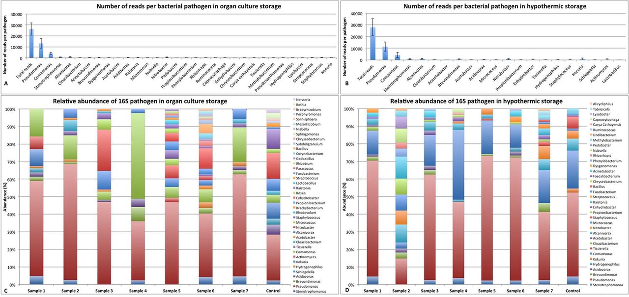
Bacterial genomic material in organ culture and hypothermic storage. (A) Cumulative number of reads per sample for 16S in organ culture and (B) in hypothermic storage. (C) Relative abundance of 16S pathogens in organ culture storage and (D) in hypothermic storage.
Sequence reads of 16S per sample in organ culture and hypothermic storage showing the presence of genomic material in the samples
Presence of fungal genomic material in OC and hypothermic storage
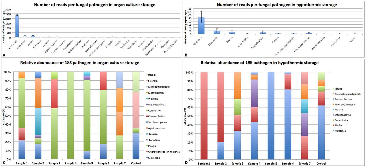
Classification of fungal organisms was assigned based on the last available taxonomy. As above, cumulative results of top 10 organisms with highest reads per sample for 18S in OC are shown in figure 2A and for hypothermic storage are shown in figure 2B. Relative abundance of fungal pathogen in OC storage is shown in figure 2C and in hypothermic storage is shown in figure 2D. Only Pinales and Malassezia spp. were found abundant both in OC and hypothermic storage. Malassezia sp. showed the highest number of reads in both the media. Most OTUs were classified from plants, mainly Malassezia, indicating that the other fungi (from plants) were absent due to the antifungal compounds present in the media. The number of reads and presence of genomic material in each sample are shown in table 3.

Fungal genomic material in organ culture and hypothermic storage. (A) Cumulative number of reads per sample for 18S in organ culture and (B) in hypothermic storage. (C) Relative abundance of fungal pathogen in organ culture storage and (D) in hypothermic storage.
Sequence reads of 18S per sample in organ culture and hypothermic storage showing the presence of genomic material in the samples
Alpha diversity of 16S and 18S in OC and hypothermic medium
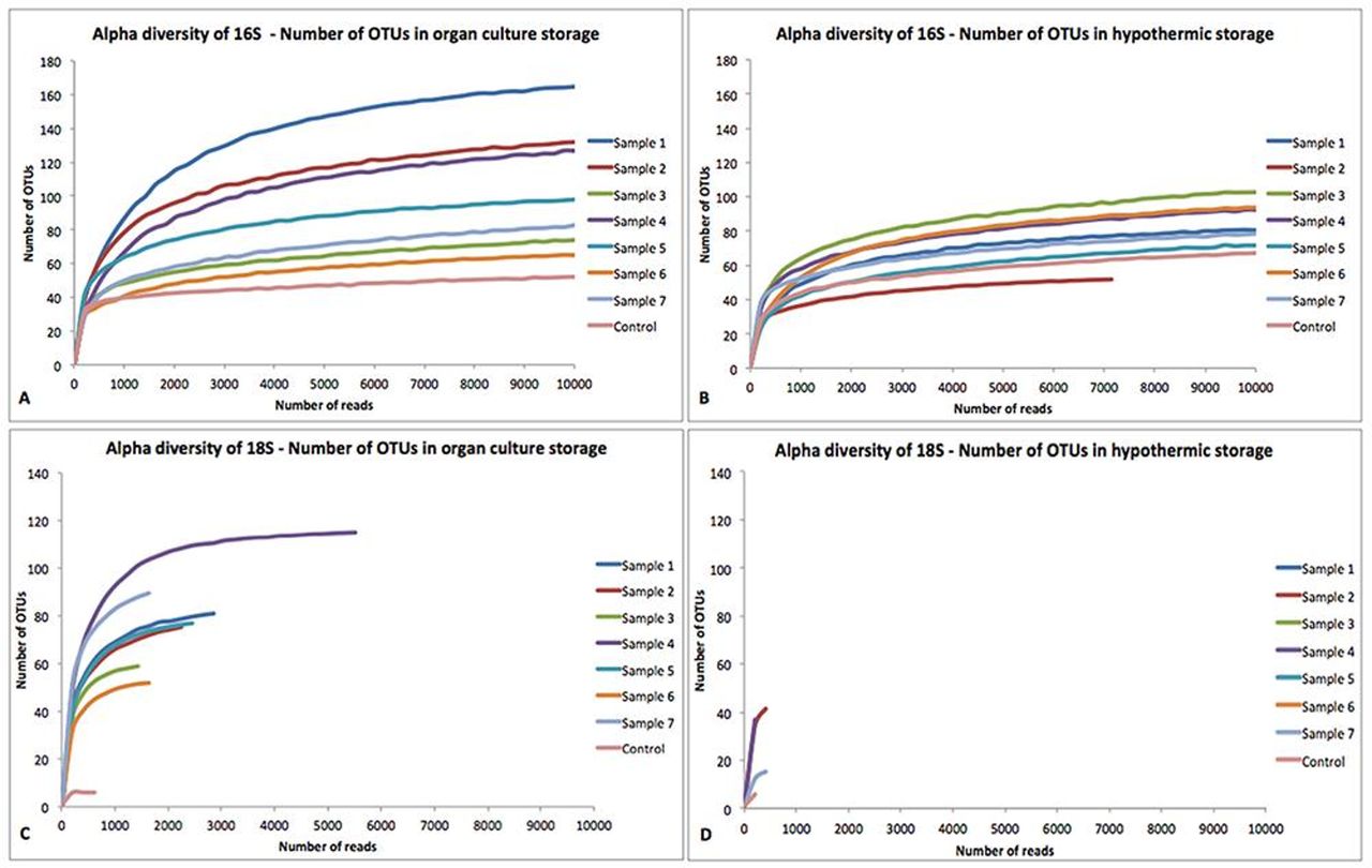
Refraction curves represent the species richness for a given number of individual samples. The number of different OTUs was plotted against the amount of reads per sample. The curves became flatter with increasing amount of reads in 16S samples indicating that only a few additional OTUs were likely to appear after deeper sequencing both, in OC (figure 3A) and in hypothermic media (figure 3B). The curves for 18S did not reach 10 000 reads both in OC (figure 3C) and in hypothermic media (figure 3D).

Alpha diversity of (A) 16S in organ culture, (B) 16S in hypothermic storage, (C) 18S in organ culture and D) 18S in hypothermic storage. OTUs, operational taxonomic unit.
Conventional diagnostic method and control group analysis
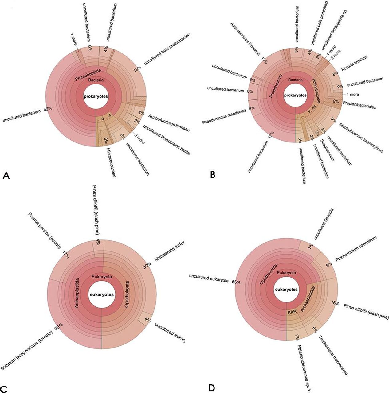
All the samples, including controls, showed negative results from CDM analysis using Bactec instrument. However, even the control samples showed presence of genomic 16S (figure 4A for OC and 4B for hypothermic media) and 18S (figure 4C for OC and 4D for hypothermic media) material that was determined as sterile by conventional microbiological assays and industrial assays.
Krona chart of control samples showing presence of genomic material in (A) 16S for organ culture and (B) for hypothermic media and (C) 18S for organ culture and (D) for hypothermic media.
Discussion
Although corneal tissue and its preservation solution should be pathogen-free before a transplant can be carried out, there are reports of corneal infection and endophthalmitis after corneal transplantation due to contaminated donor tissue.10 It is well known that traditional culture methods only detect a fraction of the available microbiota.11 Studies on the ocular surface have shown the presence of high bacterial load per 1 ng of total DNA.12 Dong et al13 reported 59 distinct bacterial genera on the ocular surface microbiome using 16S rDNA gene deep sequencing.
As there are multiple diagnostic tools available for the detection of an infection, the choice of the diagnostic method becomes important. 16S and 18S approaches are used to detect prokaryotes and eukaryotes, respectively, whereas shotgun is used for deep genome sequencing resulting into the identification and taxonomical classification of all microorganisms.14 Although 16S is useful for large number of laboratory or clinical samples, it offers limited taxonomical and functional resolution15 compared with shotgun sequencing. Shotgun sequencing can be expensive, but it has high-resolution data obtaining capacity, thereby enabling specific taxonomic and functional classification of sequences and identifying new microbial genes.
Eye banks that collect, preserve, process and distribute donated human ocular tissues, store corneas using two different approaches.16 In Europe and New Zealand, corneas are predominantly stored in an OC17 18 medium, whereas in the USA, Asia and Australia, most donor corneas are stored in short-term hypothermic conditions between 2∘C and 8∘C.16 The length of culture period (7–30 days) and the temperature (typically 31∘C–37∘C) of an OC medium facilitate the growth and detection of certain types of microorganisms.19 Endophthalmitis has been reported to occur more commonly if the donor had septicaemia.20 Septicaemia is a contraindication if the prospective donor cornea is17 21 22 stored in hypothermia. With OC, patients with bacterial septicaemia are not precluded as donors, as long as concomitant microbiological testing is performed.17 21–23 Antibacterial agents such as penicillin and streptomycin, and antifungal agents such as amphotericin B are usually used as an empiric cocktail in OC corneal preservation media. Conventional microbiological controls are currently performed using standard bacteriological media in aerobic and anaerobic environments, whereas Sabouraud broth is a routine medium for detection of fungi.24 Other options include the use of Bactec blood bottles (Becton Dickinson) incubated in the Bactec instrument (based on the detection of CO2 produced by microorganisms), which offer many advantages over the standard microbiological techniques.25–27 These techniques, however, only detect the presence of microorganisms but not their identity.
In this study, all the samples and controls showed evidence of the presence of microorganisms or its genomic content using 16S and 18S approaches. In particular, we also found same microorganisms in both, hypothermic and OC storage media. The presence of genomic material in the preservation media, however, does not necessarily relate to viable microorganisms in the storage solution. It is, therefore, not clear whether the difference between the 16S and 18S approaches and conventional culture reflects inhibition, but not eradication, of microorganisms by antimicrobials in the OC medium, differences in sensitivity and or the absence of living microorganisms or gDNA.
It is worth considering possible sources of the microbial DNA. It is possible that different genomic materials in our solutions could have come from either raw materials or packaging items when the media was manufactured. For example, genomic material of abundant microbes such as Pseudomonas, Stenotrophomonas and Comamonas spp. could have come from the industrial water. To produce highly purified water, microorganisms present in water are treated using ultrafiltration, followed by ultraviolet light that lyses the bacteria releasing genomic material into the media. The genomic material of Alcanivorax sp. could be related to the cap of the storage vial, as it is the only component that contains material derived from oil. The cap undergoes irradiation (beta or gamma), thus leading to release of genomic material. All the batches of the media were tested for 14 days in culture and the sterility in the industry is confirmed before releasing the batch. The presence of a low abundance of Brevundimonas sp. could be from the ocular surface when the corneas are cleaned with polyvinyl pyrolidone before placing them in the storage solution. It is possible that the genomic material of non-viable microorganisms may have stuck to the epithelial cells and would have been released in the storage media during preservation. Fungal (18S) contamination was at a very low abundance rate. Interestingly, OC showed a higher number of bacterial and fungal OTUs compared with that in the hypothermic media. Indicating that larger number of species could be possibly available when the conditions are optimum for the growth of an organism.
Comparing the two majorly used protocols of corneal preservation, we expected that hypothermic storage media would have less genomic material compared with OC, as OC preservation system supplements the growth condition (temperature and supplements) of microorganisms much better than hypothermic condition. The concentration of fungal DNA was higher than bacterial DNA. The absolute reads were higher in hypothermic samples compared with OC samples. As the medium is an industrial product, most of the organisms identified in our study are from the industrial raw material or water that may contain more organisms of fungal rather than of bacterial origin and therefore less bacterial DNA was observed in the samples. There are chances that such a variation could also have been due to technical issues but as all the samples were processed at the same time, this possibility could be ruled out. However, the raw materials and the final vials used for hypothermic media sampling are different than those of OC media. Some constituents or the materials could have been a possible source of more DNA concentration found from the hypothermic group compared with those from the OC group. Multiple factors such as different concentrations of antibiotics, media formulation, raw materials, downstream processing, temperature differences, etc could have also led to the presence or release of more DNA from organisms before, during or after preservation. Industrial procedures to detect live microorganisms is sufficient, but could be improved with more specific and sensitive assays like next-generation sequencing (NGS), whereas sequestered microbes in the tissue will not be detected and they have been considered to be the risk for infections such as endophthalmitis.20 Most of the DNA (regardless its provenience) came from taxa usually found in industrial water. Some of those taxa contain species that could be pathogenic. However, the amount of reads detected suggests that the actual contamination is negligible (if not just the background noise). All our samples showed negative results using Bactec colorimetric analysis, which would suggest that the samples were unlikely to contain sufficient viable microorganisms for the samples to be found positive, thus indicating that the currently used antibacterial and antifungal cocktails used in the respective media are also reliable for corneal preservation.
Aldave et al observed an insignificant increasing trend in the rate of fungal infection; they determined that it is not sufficiently compelling to pursue antifungal supplementation for donor storage media.28 In this study, we also report that fungal contaminants were found at a very low abundance rate. The other microorganisms detected that might have arisen from the cornea or the media may have been below the detection limit of CDC or were killed by the antimicrobials present inside the media. The 10 most abundant genera found on the ocular surface include Pseudomonas, Propionibacterium, Bradyrhizobium, Corynebacterium, Acinetobacter, Brevundimonas, Staphylococcus, Aquabacterium, Sphingomonas and Streptococcus,29 which were also observed in our samples.
16S and 18S data were acquired and analysed, which only provides data on the detection of genes and not necessarily viable microorganisms, which could be considered as a potential limitation of this study. This could, however, be supplemented with proteomics to detect live organisms. The other limitation is that the method measures only rRNA and therefore other genomic information is missing and specificity of identification is reduced. By law, if the storage media is contaminated, the corneal samples must be discarded. With further improvements, NGS could be advantageous by detecting the presence of genomic material in a short span of time and with reduced costs. A controlled comparative in vitro study of NGS and CDC with enrichment culture and removal of antibiotics in the medium is needed.
Current study showed the presence of gDNA in the negative control samples. A positive control of a known organism and concentration would have been beneficial for understanding the efficiency and sensitivity of metagenomics. Because of the high sensitivity of this technique, technicians must strictly follow a total sterility protocol avoiding contamination during sample processing. The cornea sheds epithelial cells during the preservation phase. Regeneration of these cells in OC particular, if co-infected by intracellular microorganisms, highlights the need for their detection by NGS especially as it has been observed that ocular surface contains a small amount of bacterial cells.
Metagenomic deep sequencing has the potential to improve the microbiological analysis of samples starting from low concentrations. The costs, presence of live organisms, turnover time, downstream processing and data analysis could be considered as limitations when it comes to routine eye banking procedures especially when the empiric solutions already seem to be relatively safe. Given the current trends in genomic technology development, the costs are likely to be reduced significantly and more narrowed and standardised results will be obtained in the near future. Wilson et al showed that with adequate staffing, the final protocol could be completed in less than 48 hours.30 NGS could therefore be of significant value for checking the microbiological load in industrial production to ensure the safety of healthcare products. Metagenomics has a role for detecting organisms with high specificity and sensitivity, which may also be important at the centres where Good Manufacturing Practices (GMP) rules are stringent.
References
Footnotes
MP and DB contributed equally.
Contributors Concept and design of the study: MP, DB, DC, SF. Drafting manuscript: MP, DB, SBK, SF. Critical revision of manuscript: MP, DB, VR, SBK, DC, DP, SF. Supervision: SBK, DC, DP, SF. Final approval: MP, DB, VR, SBK, DC, DP, SF.
Funding 2017 Camera di Commercio di Venezia Rovigo Delta Lagunare funds. CUP I78C17000040005. 5X1000 funds (year 2016) to Fondazione Banca degli Occhi del Veneto Onlus (Venice, Italy) by the Italian Ministry of Health and Italian Ministry of University and Research.
Competing interests None declared.
Patient consent for publication Not required.
Provenance and peer review Not commissioned; externally peer reviewed.