Article Text

Abstract
Aims To examine the expression of Toll-like receptor (TLR) 5 in the conjunctival epithelium of patients with severe ocular surface diseases.
Methods Immunohistochemical study of TLR5 was performed on conjunctival tissues obtained from patients undergoing surgical reconstruction of the ocular surface to treat Stevens-Johnson syndrome (SJS) (n=4), ocular cicatricial pemphigoid (OCP) (n=3), chemical eye burn (n=3), and pterygium (n=2), and on nearly normal conjunctival tissues obtained during surgery for four cases of conjunctivochalasis as a control.
Results TLR5 protein was consistently and abundantly expressed in the conjunctival epithelium and detected only at the basal and wing cells. However, in the conjunctival epithelium obtained from the patients with SJS, OCP and chemical eye burns, the TLR5 protein was detected at not only the basal and wing cells but also at the superficial cells. TLR5 protein detected in the pterygium patients mirrored that detected in the controls.
Conclusions Although TLR5 was normally present on the basal and wing cells of conjunctival epithelium with spatially selective presence, it was expressed on not only the basal and wing cells but also the superficial cells in the conjunctival epithelium of patients with SJS, OCP or chemical eye burns, suggesting that TLR5 might be upregulated in the conjunctival epithelium of these diseases.
- Ocular surface
- Inflammation
- Conjunctiva
- Cornea
Statistics from Altmetric.com
Introduction
The ocular surface epithelium serves as the defensive front line of the innate immune system, and Toll-like receptors (TLRs) are known to be one of the key receptors of the innate immune system. Reportedly, TLRs are pattern-recognition receptors that sense conserved pathogen-associated molecular patterns (PAMPs), and are the key receptors for the recognition of microbes.1 TLRs are a type of transmembrane protein composed of three major domains, and are characterised by LRRs (leucine-rich repeats) in the ectodomain which mediate the recognition of their respective PAMPs. TLR5 recognises bacterial flagellin, a component protein of bacterial flagella.2 ,3 Flagella are present in both gram-positive and gram-negative bacteria and are essential for bacterial motility, invasion and chemotaxis.4 We previously reported that human ocular surface epithelial cells, both corneal and conjunctival, express TLR5-specific mRNA and TLR5 proteins, and TLR5 proteins were detected at basal and wing site cells.5 ,6 The purpose of this present study was to examine the expression of TLR5 in the conjunctival epithelium of patients with severe ocular surface disorders.
Methods
This study was approved by the Institutional Review Board of Kyoto Prefectural University of Medicine, Kyoto, Japan, and all experiments were conducted in accordance with the tenets set forth in the Declaration of Helsinki. The purpose of the research and the experimental protocol were explained to all patients, and their informed consent was obtained before their involvement in the study.
For the immunohistochemical study, the control samples were nearly normal human conjunctival tissues obtained at the time of surgery for the treatment of conjunctivochalasis (four cases). Conjunctivochalasis is a normal ageing related change, and it is an isolated bilateral condition in which redundant bulbar conjunctival tissue interposes between the globe and the lower eyelid.7 Cicatricial conjunctival tissues were also obtained from patients undergoing surgical reconstruction of their ocular surface that had been devastated due to Stevens-Johnson syndrome (SJS) (four cases), ocular cicatricial pemphigoid (OCP) (three cases), and chemical eye burns (three cases). In addition, the conjunctival tissues of patients with pterygium (two cases) were obtained. Pterygium is reportedly a benign growth of the conjunctiva that is thought to be caused by exposure to ultraviolet light.8
For TLR5 staining, we used mouse antihuman TLR5 monoclonal antibody (mAb; Abcam, Cambridge, UK) (TLR5: TLR5 antibody (19D759.2)) in a 0.5 μg/μL dilution with blocking solution. As isotype controls, mouse IgG2a X0943 (Dako Cytomation, Kyoto, Japan) in a 0.1 μg/μL dilution was used. After incubation overnight at 4°C in a moist chamber, the sections were thoroughly washed with 0.01 M phosphate buffered saline. Next, the sections were subjected to secondary antibody reactions with biotin conjugated donkey anti-mouse IgG Fab fragments in a 0.5 μg/μL dilution. Avidin and biotinylated horseradish peroxidase macromolecular complex reagent (VECTASTAIN ABC reagent; Vector Laboratories, Inc, Burlingame, California, USA) was then applied to the section for 30 min and 3,3′-diaminobenzidine peroxidase substrate solution (DAB substrate kit; Vector Laboratories) was added to the slide sections as a chromogenic substrate. Finally, the sections were counterstained using haematoxylin.
Results
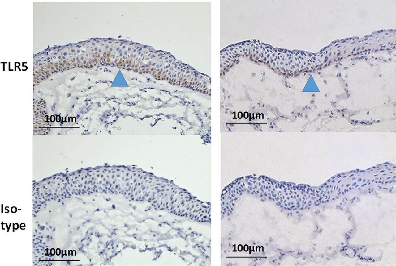
Conjunctival tissues obtained at the time of ocular surface reconstruction surgery were subjected to immunohistochemical study to determine the presence and localisation of TLR5 expression in stratified conjunctival epithelium. In human conjunctival epithelium of the control conjunctiva, TLR5 protein was consistently and abundantly expressed; however, it was detected only at the basal and wing cells (figure 1). These results are consistent with those of our previous report.6 On the other hand, in the epithelium of the conjunctival tissues obtained from the four SJS patients (figure 2A), the three OCP patients (figure 2B), and the three patients being treated for chemical eye burns (figure 2C), the TLR5 protein was detected in all layers from the basal and wing cells to the superficial cells. However, in the epithelium of the conjunctival tissues obtained from the two pterygium patients (figure 3), the TLR5 protein was detected in its basal and wing cells, but not the superficial cells, the same as with the controls.

Immunohistological analysis of Toll-like receptor (TLR) 5 in the conjunctival epithelium of the controls. Localisation of TLR5 in human conjunctival epithelium as control tissue, from four conjunctivochalasis cases which underwent surgery. The conjunctival tissue was incubated with anti-TLR5 antibody. Isotype control incubation was used as the negative control.

Immunohistological analysis of Toll-like receptor (TLR) 5 in the conjunctival epithelium of various ocular surface disorders. Localisation of TLR5 in the conjunctival epithelium of various conjunctival disorders; four Stevens-Johnson syndrome cases in the chronic stage (A), three ocular cicatricial pemphigoid cases (B), and three chemical burn cases (C). The conjunctival tissue was incubated with anti-TLR5 antibody. Isotype control incubation was used as the negative control.

Immunohistological analysis of Toll-like receptor (TLR) 5 in the conjunctival epithelium obtained from two pterygium patients. Localisation of TLR5 in the conjunctival epithelium from the two pterygium cases which underwent surgery. The TLR5 protein was detected only at the basal and wing sites, the same as with the control. Isotype control incubation was used as the negative control.
Discussion
The findings of this study showed that in devastating ocular surface disorders such as SJS, OCP and chemical eye burns, TLR5 is expressed not only on the basal and wing cells but also on the superficial cells, although TLR5 is expressed on only the basal and wing cells in normal human conjunctival epithelium as we previously reported.6 In the conjunctival epithelium obtained from the two pterygium patients, TLR5 protein was also consistently and abundantly expressed, yet detected only at the basal and wing cells, the same as with normal human conjunctival epithelium. The above findings suggest that TLR5 might be upregulated in the ocular surface of patients with these devastating ocular surface disorders.
It should be noted that previous reports have investigated the expression of TLR5 in other human disorders. Recent findings have revealed that both in rheumatoid arthritis (RA) and osteoarthritis, TLR5 immunostaining is significantly higher on synovial tissue lining, sublining macrophages and endothelial cells compared with controls.9 The findings of that study also suggest that the expression of TLR5 is upregulated in RA disease progression, that RA is reportedly a chronic autoimmune disorder in which the innate immune system plays an important role, and that intestinal epithelium reportedly expresses TLR5.
Rhee et al10 reported that the flagellin/TLR5 response is confined to the basal cells, not the superficial cells, of intestinal mucosa, indicating that the intact colonic mucosa is not responsive to luminal bacterial flagellin. We also reported that on ocular surface epithelium, TLR5 was expressed on only the basal and wing cells, not the superficial cells, thus suggesting that in the normal condition, the intact ocular surface could not be responsive to commensal bacterium of the ocular surface.11 In a very recent study, it was reported that TLR5 mRNA tends to be upregulated in the active phase of ulcerative colitis (UC) compared to UC quiescent disease, and that there are positive correlations between TLR5 mRNA and endoscopic and histological activity.12 Moreover, in irritable bowel syndrome (IBS) of dysfunctional colitis that partly results from low grade mucosal inflammation, it was reported that TLR5 mRNA was upregulated in colonic biopsies from active IBS patients.13 Therefore, TLR5 expressions may be upregulated in the diseases associated with chronic inflammations or in an active inflammation phase.
In chronic phase SJS and OCP patients, it was reported that the number of neutrophil elastase-positive cells and CD4-positive cells was increased in the epithelium and substantia propria of the pannus tissue. In addition, CD3-positive cells in the substantia propria of the pannus were reportedly slightly increased in both OCP and SJS patients, yet other kinds of infiltrating cells were not increased.14 Therefore, upregulation of TLR5 in the epithelium of the conjunctival tissues of SJS, OCP, and chemical eye burns might be involved in the chronic inflammation of these respective diseases. Although it has been reported that there are chronic inflammatory cells in pterygium,15 the difference of the extent of TLR5 expression between pterygium and the devastating ocular surface disorders might suggest the different quality of the chronic inflammation.
Acknowledgments
The authors wish to thank John Bush for reviewing the manuscript.
References
Footnotes
-
Contributors Writing the manuscript: MU and KY. Research design and obtaining funding: MU. Provision of materials: CS, NY, TI and SK. Literature search: KY.
-
Funding This work was supported by Grant in Aids from Ministry of Education, Culture, Sports, Science and Technology of the Japanese government (no.25293355), and also partly supported by grants-in-aids for scientific research from the Japanese Ministry of Health, Labour and Welfare, and a research grant from the Kyoto Foundation for the Promotion of Medical Science and the Intramural Research Fund of Kyoto Prefectural University of Medicine.
-
Competing interests None.
-
Patient consent Obtained.
-
Ethics approval The Institutional Review Board of Kyoto Prefectural University of Medicine, Kyoto, Japan.
-
Provenance and peer review Not commissioned; externally peer reviewed.