Article Text

Abstract
Background In vivo retinal imaging of rodents has gained a growing interest in ophthalmology and neurology. The bedding of the animals with the possibility to perform adjustments in order to obtain an ideal camera-to-eye angle is challenging.
Methods We provide a guide for a cost-effective, do-it-yourself rodent holder for ocular imaging techniques. The set-up was tested and refined in over 2000 optical coherence tomography measurements of mice and rats.
Results The recommended material is very affordable, readily available and easily assembled. The holder can be adapted to both mice and rats. A custom-made mouthpiece is provided for the use of inhalant anaesthesia. The holder is highly functional and assures that the rodent’s eye is the centre of rotation for adjustments in both the axial and the transverse planes with a major time benefit over unrestrained positioning of the rodents.
Conclusion We believe this guide is very useful for eye researchers focusing on in vivo retinal imaging in rodents as it significantly reduces examination times for ocular imaging.
- holder
- rodent
- ocular imaging
- do-it-yourself
This is an Open Access article distributed in accordance with the Creative Commons Attribution Non Commercial (CC BY-NC 4.0) license, which permits others to distribute, remix, adapt, build upon this work non-commercially, and license their derivative works on different terms, provided the original work is properly cited and the use is non-commercial. See: http://creativecommons.org/licenses/by-nc/4.0/
Statistics from Altmetric.com
Key messages
While in vivo ocular imaging of small rodents, namely optical coherence tomography (OCT), is gaining increasing importance in ophthalmology and neurology, the available devices often require additional holders to manipulate the animals, especially if different species are to be imaged.
We provide a do-it-yourself guide for a new whole-body positional manipulator for ocular imaging of mice and rats. It is cost-effective, easily assembled and compatible with any OCT device on the market.
The holder speeds up OCT and fundus imaging in rodents and enables inhalation anaesthesia, which increases the throughput and reduces the recovery time from anaesthesia.
BACKGROUND
In recent years, in vivo retinal imaging has gained increasing relevance1–3 not only in ophthalmological4–8 but also in neurological9–19 cases. Optical coherence tomography (OCT) and confocal scanning laser ophthalmoscopy (cSLO) have been identified as useful diagnostic tools to evaluate a large variety of retinopathies and retinal manifestations of neurological diseases. OCT allows for fast, non-invasive and high resolution in vivo visualisation of the retinal morphology and has been introduced as an outcome parameter in clinical trials of neuroprotection in multiple sclerosis and optic neuritis.20–22
The high resolution of third-generation spectral-domain OCT devices renders in vivo retinal imaging in mice and rats possible, gaining an increasing importance in ophthalmological and neurological preclinical research.23–33 The obtained results are in good accordance with histological sections of the animals’ retinae.34
The application of OCT technology in rodent models, however, is still challenging, mainly because of the small size of the rodents’ eyes. Even if systems have been developed specifically for the imaging of rodents,31 35 several commercially available devices require adaptations to image animals of different species. If investigators desire to change the species under investigation, for example, mice to rats, often a different size of holder is required. Animals have to be anaesthetised for measurement, which is largely facilitated by the proposed mouthpiece and holder.36 37 Positioning the rodent with the option to easily adjust the eye orientation is critical for reproducible and high-quality images. Different holders have been proposed38–43 to assure an optimal positioning and an adequate camera-to-eye angle. However, not all holders are applicable with all OCT devices. Some holders do not harmonise with the size of the OCT camera on certain devices, some are patented and/or only available together with OCT devices and most are high priced. We, therefore, aimed to develop a guide and instructional drawing for a do-it-yourself, cost-effective and adjustable holder for bedding of rodents during OCT and cSLO measurements as well as for other ocular imaging techniques. The device is in use at our facilities and delivers excellent results with rodent OCT imaging.
METHODS
Ethics
The set-up was tested and approved in over 2000 measurements with mice and rats. All animal procedures were performed in compliance with the experimental guidelines approved by the regional authorities (State Agency for Nature, Environment and Consumer Protection; AZ 84-02.4.2014.A059) and conform to the Association for Research in Vision and Ophthalmology Statement for the Use of Animals in Ophthalmic and Vision Research.
Material
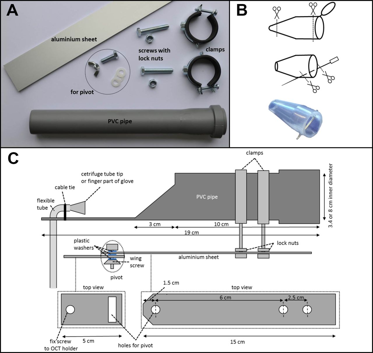
For the construction of the holder, we recommend the following items (figure 1A):

Construction and properties of the holder. (A) Recommended items for the holder, (B) construction of the mouthpiece and (C) diagram for modification of the items and assembly of the holder. OCT, ocular coherence tomography.
Polyvinylchloride (PVC) pipe, inner diameter: ~3.4 cm for mice, ~6–8 cm for rats (eg, standard drainage pipes), approximately US$3;
Aluminium sheet (20 × 5 × 0.2 cm), approximately US$11;
Screw (M8, 4 cm), wing screw and plastic washers for the pivot, approximately US$1;
Two clamps (size matching the PVC pipe, eg, drainage pipe holders), screws and lock nuts, approximately US$5.
Tools
The following tools are needed for the construction of the holder:
jigsaw (saw blade for metal and plastic)
drill (bits for metal)
file
screw wrenches.
Assembly of the holder for mice
The following is a guide for the assembly of a holder for mice. Adaptations necessary for a rat holder are described below. Shorten the PVC pipe to a final length of 19 cm. At 10 cm from one end, cut out the top of the tube in a sloping shape, leaving a 6 cm long and 3 cm wide strip as illustrated in figure 1C. Drill a hole of 0.8 cm diameter into the strip at the front end of the PVC pipe (~2 cm from the front end) to hold the tube for the inhalant anaesthesia supply. Cut the aluminium sheet into two pieces of 15 and 5 cm length. Drill a rectangular excision of 1 x× 4 cm at 1 cm from the end of the smaller sheet for a slidable pivot and an additional circular hole for an M8 screw at 0.5 cm from the other end. Slope the tip of one end of the 15 cm aluminium sheet at a 45°° angle using the jigsaw (optional) and drill three M8 holes at 1.5, 7.5 and 10 cm from the sloped end. Assemble the items by inserting the plastic washers between the screw, the two aluminium sheets and the wing screw for smooth movement of the pivot. The tube for inhalant anaesthesia supply (if needed) can be inserted and fixed with a cable tie. To form a mouth piece with an integrated bite bar, cut a centrifuge tube tip at 0.8 and 2.4 cm from the narrow end. Pierce a 20-gauge syringe needle through the bottom third of the tube tip at 0.3 cm from the broad end (figure 1B). This mouthpiece design allows a snug fit for the mouth of adult animals of most commercially available mouse lines (eg, A/J, BALB/cJ, C57BL/6J, DBA/2J, NMRI, SJL) and provides an improved immobilisation of the animal by carefully hooking the upper front teeth over the bite bar. Anaesthesia can be maintained by inhalation of isoflurane vaporised with pure oxygen at concentrations of 2% during measurement.
To maintain the body temperature and ensure immobility, we suggest wrapping the animal in a paper towel during measurement. Additionally, an external heat source, for example, a heat string used for terrariums, can be wrapped around the holder for longer measurements.
Adaptations for a holder suitable for rats
For rat holders (eg, Sprague Dawley, Wistar, Lewis, Long Evans, Brown Norway), the PVC pipe should have an inner diameter of ~6-–8 cm with size-matching clamps. The inhalant anaesthesia supply tube can be modified using the finger part of a laboratory glove, cut open on both sides and fixed on the tube with tape.
OCT device and fixation of the holder
Our experiments were performed using a Heidelberg Engineering Spectralis HRA+OCT device (Heidelberg Engineering GmbH, Heidelberg, Germany). The holder was attached to the platform that holds the chin rest for human subjects and that can be adjusted in the z-axis. However, the device is compatible with any other OCT device on the market and can be attached to a simple xyz table.
Statistics
A paired Student’s t-test was performed to compare the time needed to adjust the mouse’s eye position for an OCT measurement with and without the use of the rodent holder. Average values are presented as mean with SD. Differences were considered significant at p<0.05.
RESULTS
Evaluation of benefits for retinal imaging
Analysing the time needed to achieve correct positioning of the eye-to-camera angle and scanning area in OCT measurements of 11 independent mouse eyes, we observed a highly significant (p<0.001) time benefit when using the rodent holder (figure 2). The time expended was almost three times higher (2.8×) when the animal was manually positioned and the angles of imaging were adjusted by moving the OCT camera.

Time to adjust the mouse's optic disc in a centric position for optical coherence tomography imaging with and without the rodent holder. Boxes and whiskers represent the mean, SD and minimum to maximum of results obtained while imaging 11 independent eyes (***p >0.001, Student's t-test).
Furthermore, in our experience, the use of the holder seems to reduce the movement artefacts from breathing; however, this was not formally assessed.
DISCUSSION
For in vivo ocular imaging, especially retinal OCT and fundus imaging, a beam path through the middle of the pupil with an orthogonal angle to the target structure (eg, the retina) in all planes is an essential prerequisite for optimal image quality and reproducibility in longitudinal assessments. During live imaging, this can be obtained by modifying the angle of the camera, of the animal or both. Cameras for retinal imaging are often heavy, and the design of camera holders allowing movements in all planes and angles is challenging. Some OCT devices only allow movements in the vertical axis, so the adjustment of the angle as well as the horizontal and vertical axes has to be performed by moving the animal. An advantage of adjusting the beam-to-target angle by moving the animal is that the camera can be manoeuvred in the z-axis without changing the angle of imaging. This is not possible when the camera is moved from an angled position.
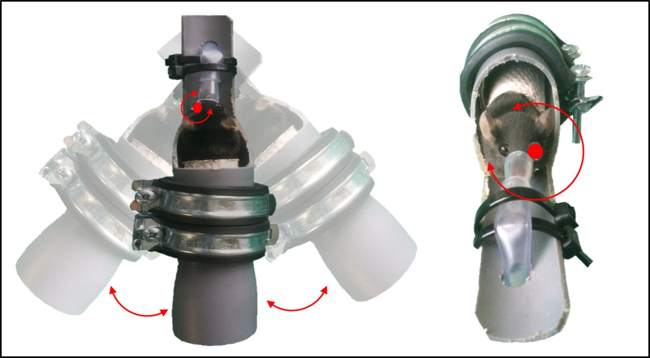
A remarkable strength of the proposed holder is that the rodent’s eye is the centre of rotation for rotations in both the axial and transverse planes (figure 3). By adjusting the position of the mouth piece that tightly holds the rodent’s head by the upper front teeth, which can be hooked over the bite bar, the eye can be positioned in the centre of the rotational pivot of the aluminium sheets and in the middle of the tube. Therefore, the beam-to-target angle can be adjusted by a manual rotation of the holder tube housing the animal. In most OCT systems, the camera can be moved along the visual axis of the camera to zoom into the eye and provide adjustments of the z-axis. Movements in the x-plane and y-plane can be performed using the pivot of the aluminium sheets of the holder, which is also slidable due to the rectangular excision at the end of the smaller aluminium sheet. If additional movements are needed (ie, if the camera cannot be moved in the z-axis), the holder can be fixed on a simple xyz table. Our experiments demonstrate that this reduces the time needed to find the correct angle in all planes compared with angling of the camera. Considering an average imaging time of 2 min for high-resolution volume scans per eye, the time benefit of 30 s corresponds to a reduction of time needed for imaging by 25%. However, we have to acknowledge that, with the experience and training, imaging can also be performed without the use of a holder by just placing the animals on a stable platform if the camera can be moved and rotated in all angles and planes and operators may become quicker in adjusting the animal with practice. Overall, in our experience, the use of the holder has largely facilitated rodent imaging, reducing acquisition times and served to avoid having to readjust the animals or repeat the measurements due to movement or dislocation of the animal.
Rotational axis around the rodent eye. Rotation in transverse plane (left) and in axial plane (right).
Another advantage of the device is that it includes an option for volatile anaesthesia. The combination of fixation of the mouth using the bite bar and volatile anaesthesia serves to reduce breathing artefacts during image acquisition. Furthermore in our experience, volatile anaesthesia (eg, inhalant isoflurane) is safer and easier to control than injectable anaesthesia (eg, ketamine-–xylazine),44 45 and serves to prevent premature awakening of rodents in case of longer acquisition times. This allows imaging for over 45 min, for example, for funduscopic imaging of fluorescence-labelled cells or complex OCT protocols. However, the combination of ketamine-–xylazine is also an effective anaesthesia method for small laboratory animals23 34 and in our experience, we have never encountered cataracts or corneal alterations during the first anaesthesia when performing OCT imaging. The method is safe, especially when using low dosages of xylazine (5 mg/kg).
We acknowledge that our holder does show resemblance to other commercially available options. This is owing to the fact that there are only a few ways to design a holder, which allows pivotal rotation of the rodent in two planes with the eye as centre of rotation. Our aim was not to develop an entirely new concept but rather to provide a do-it-yourself guide for researchers to construct a very cost-efficient yet fully functional holder. This will be of greatest help not only for research groups beginning mouse retinal imaging but also for groups planning to extend their species of interest from mice to rats. Of note, the proposed guide has been developed over a period of 12 months while performing more than 2000 retinal imaging sessions on rodents. During this period, several adaptations have been evaluated iteratively. Options such as stereotactic holders fixing the mice at the mouth or ears have proven to have no advantage in our hands over the proposed breathing mask with the bite bar and were therefore discarded. In our experience, the holder outperforms commercial options, which are available for more than 10 times the price.
A limitation of the proposed holder is that the rotations are performed manually instead of controlled dials, which may reduce the precision of very fine adjustments. In our experience, however, the movements needed for setting the angle for OCT and fundus imaging are not too delicate and can be performed manually at a sufficiently high precision. Furthermore, the proposed guide is not intended as a rigid instruction manual but rather as a prototype for researchers to introduce further refinements, including using higher quality materials, according to their specific needs and interests.
CONCLUSION
In conclusion, we present a do-it-yourself guide for a highly functional and effective rodent holder using materials that are both easily available and very affordable. Using the device speeds up OCT and fundus imaging in rodents and enables inhalation anaesthesia. Together, this increases the throughput and reduces the recovery time from anaesthesia. We believe this guide could be very useful for eye researchers aiming to establish retinal imaging in rodents at their laboratories.
References
Footnotes
Contributors MD made substantial contributions to the conception and design of the holder, acquisition of data as well as analysis and interpretation of data; he has been involved in drafting and writing the manuscript. AC-H and HY made substantial contributions to the conception and design of the holder and have been involved in drafting the manuscript. AB and HPH have been involved in revising the manuscript critically for important intellectual content and made substantial contributions to the interpretation of data. OA has been involved in revising the manuscript critically for important intellectual content. AG made substantial contributions to the conception and design of the holder and has been involved in revising the manuscript critically for important intellectual content and made substantial contributions to the interpretation of data. PA made substantial contributions to conception and design of the holder, acquisition of data as well as analysis and interpretation of data; he has been involved in drafting and writing the manuscript, and has given final approval of the version to be published and agreed to be accountable for all aspects of the work in ensuring that questions related to the accuracy or integrity of any part of the work are appropriately investigated and resolved.
Funding The rodent holder was developed and refined in the context of research projects that were funded by grants from Dr. Robert Pfleger Stiftung, Forschungskommission der Heinrich Heine University Düsseldorf, Novartis and Biogen to PA.
Competing interests None declared.