Article Text

Abstract
Objective To describe the clinicopathological and genomic features of nine patients with primary and secondary orbital/ocular manifestations of leukaemia.
Methods All orbital/ocular leukaemic specimens from 1980 to 2009 were collected from the Danish Register of Pathology. In six cases, medical records and formalin-fixed, paraffin-embedded blocks were available. Three cases from the Department of Pathology, Royal Liverpool University Hospital, were also included. Immunophenotypes and MYB oncoprotein expression were ascertained by immunohistochemistry. Genomic imbalances were analysed with comparative genomic hybridisation arrays and oncogene rearrangements with fluorescence in situ hybridisation.
Results Four patients had B-cell precursor acute lymphoblastic leukaemia (BCP-ALL) and five had acute myeloid leukaemia (AML). Two patients with BCP-ALL and one with AML had primary orbital manifestations of leukaemia. Common symptoms were proptosis, displacement of the eye, and reduced eye mobility in patients with orbital leukaemias and pain, and reduced visual acuity in patients with ocular leukaemias. All patients with primary orbital lesions were alive up to 18 years after diagnosis. All but one patient with secondary ophthalmic manifestations died of relapse/disseminated disease. ETV6 and RUNX1 were rearranged in BCP-ALL, and RUNX1 and KMT2A in AML. Genomic profiling revealed quiet genomes (0–7 aberrations/case). The MYB oncoprotein was overexpressed in the majority of cases.
Conclusions Leukaemias with and without ophthalmic manifestations have similar immunophenotypes, translocations/gene fusions and copy number alterations. Awareness of the clinical spectrum of leukaemic lesions of the eye or ocular region is important to quickly establish the correct diagnosis and commence prompt treatment.
- acute leukaemia
- ophthalmic manifestations
- ocular lesions
- clinical characteristics
- gene fusion
- array comparative genomic hybridization
This is an open access article distributed in accordance with the Creative Commons Attribution Non Commercial (CC BY-NC 4.0) license, which permits others to distribute, remix, adapt, build upon this work non-commercially, and license their derivative works on different terms, provided the original work is properly cited, appropriate credit is given, any changes made indicated, and the use is non-commercial. See: http://creativecommons.org/licenses/by-nc/4.0/.
Statistics from Altmetric.com
- acute leukaemia
- ophthalmic manifestations
- ocular lesions
- clinical characteristics
- gene fusion
- array comparative genomic hybridization
Key message
What is already known about this subject?
Leukaemic infiltrates may occur in the eye or ocular region, and in rare cases it is the first presenting symptom of leukaemia.
Leukaemias with ophthalmic manifestations are rarely biopsied or examined extensively, and therefore our knowledge about these lesions is limited.
What are the new findings?
This is the first comprehensive, integrated clinicopathological, cytogenetic and genomic analyses of acute leukaemias with ophthalmic manifestations.
We show for the first time that leukaemias with eye and/or ocular manifestations have similar genetic and molecular profiles compared with other non–site-specific leukaemias.
How might these results change the focus of research or clinical practice?
Our findings further emphasise the broad clinical spectrum of leukaemic lesions with ophthalmic manifestations and the need to consider leukaemia in the differential diagnosis of patients with proptosis, reduced mobility and/or displacement of the eye.
Awareness of the clinical spectrum of leukaemic lesions of the eye or ocular region is important to quickly establish the correct diagnosis and commence prompt treatment.
Introduction
Leukaemic infiltrates can occur in the eye or ocular region as a primary manifestation of leukaemia or as an infiltration of systemic disease.1 2 Ophthalmic manifestations are more common in acute leukaemias than in chronic leukaemias.1 3 Typical symptoms of patients with leukaemic infiltrates in the ophthalmic region include eyelid oedema and swellings, chemosis and exophthalmos. Globe displacement by a leukaemic mass may restrict ocular mobility and in some cases reduce visual acuity.4 Orbital tumours may present as a diffuse infiltrate or as a large single mass. Solid infiltrates can involve all structures in the extraocular region, including the ocular adnexa and the optic nerve,4–6 and constitute approximately 2% of all malignancies in this region.5
Intraocular manifestations of leukaemias are most frequently seen clinically in the retina, but on histopathological examination, leukaemic lesions of the choroid are equally as common.4 6 Patients with intraocular leukaemic manifestations often have reduced vision, and occasionally also pain or decreased mobility of the eye. Retinal detachment, chemosis, retinitis, glaucoma, uveitis or hypopyon are observed on clinical examination.4 6
Ophthalmic signs and symptoms may also result from side effects associated with leukaemia, such as anaemia, thrombocytopenia, hyperviscosity, immunosuppression and infections.1 4 6 When a primary leukaemia arises in the eye or ocular adnexa, subsequent involvement of the peripheral blood or bone marrow usually occurs within 1 year of the ocular disease.7
Leukaemias with ophthalmic manifestations are rarely biopsied or examined extensively,1 5 and therefore our knowledge about these lesions is still limited. In this study, we describe in detail the clinical, cytogenetic, and genomic features of nine cases of primary and secondary ophthalmic leukaemias.
Materials and methods
Patient material
All cases of ocular and ocular adnexal leukaemic lesions from 1980 to 2009 were collected from the Danish Register of Pathology. In six cases, medical records and formalin-fixed, paraffin-embedded (FFPE) tissue blocks were available. Also included were three leukaemias with ophthalmic manifestations from the Department of Pathology, Royal Liverpool University Hospital, UK. The following information was collected from the patients’ medical records: age, sex, diagnosis, histopathological findings, cytogenetic data, molecular genetic data (when available), location of the lesion, symptoms, treatment and clinical follow-up.
Histopathology and immunohistochemistry
The orbital and ocular biopsies were evaluated on sections stained with H&E and periodic acid-Schiff. For immunohistochemistry, FFPE tissue blocks were cut into sections 4 µm thick and mounted on slides. Stains were performed using the streptavidin–biotin method. Antibodies against CD3, CD10, CD13, CD15, CD20, CD33, CD34, CD43, CD45, CD79α, CD117, MPO, TLC1, BCL-2, TdT and lysozyme were used in most cases. The expression of the MYB oncoprotein (SPM-175; Santa-Cruz, Dallas, Texas, USA) was studied as described.8 Samples were scored as MYB positive when >25% of the neoplastic cells stained positive for MYB. Negative and positive controls were included in all stains, and internal positive controls were evaluated where appropriate. For analyses of immunostainings, positive tumour cells were counted in five high-power fields (×400).
Array comparative genomic hybridisation (arrayCGH) analysis
Genomic DNA from FFPE blocks was isolated with the DNeasy Blood and Tissue Kit (Qiagen, Hilden, Germany). In seven cases, there was enough DNA available for arrayCGH analysis with Human Genome CGH Microarray 244K oligonucleotide arrays (Agilent Technologies, Santa Clara, California, USA).8 Data were analysed with NEXUS Copy Number V.7.0 Discovery Edition (BioDiscovery, El Segundo, California, USA).8 The settings for aberration calls were 1.5 for amplification, 0.3 for gain, –0.3 for loss and –1.5 for homozygous deletion. The FASST2 segmentation algorithm was used to define non-random regions of copy number alterations (CNAs) across the genome at a significance threshold of p=1.0E−8. In samples from cases 2 and 5, the settings for aberration calls were 1.5 for amplification, 0.5 for gain, –0.5 for loss and –1.5 for homozygous deletion at a significance threshold of p=1.0E−18. The accuracy of each aberration call was confirmed manually.
Fluorescence in situ hybridization (FISH)
Rearrangements of ETV6 and KMT2A were analysed on 5 µm FFPE sections with FISH dual-colour break probes (Leica Biosystems, Wetzlar, Germany). The protocols for pre-treatment, hybridisation and post-hybridisation washes were as recommended by the manufacturer. Fluorescence signals were digitised, processed and analysed with the Isis FISH imaging system V.5.5 (MetaSystems, Altlussheim, Germany). At least 50 nuclei were scored for each probe and case.
Patient and public involvement
Patients and the public were not involved in the design, conduct and reporting of the research. However, permission was obtained to include photographs of two of the patients in the publication.
Results
Clinical characteristics of primary ophthalmic leukaemias
We identified three cases of acute leukaemias with primary ophthalmic manifestations in the Danish Register of Pathology from 1980 to 2009. The clinical, cytogenetic and molecular genetic findings are summarised in table 1.
Clinical and cytogenetic findings and gene rearrangements/mutations in nine cases of acute leukaemia with ophthalmic manifestations
Case 1 was an otherwise healthy 5-year-old boy with left-sided exophthalmos, a swollen lacrimal gland and a bluish-red discolouration of the eyelid. The eye examination, including visual acuity, was normal. A CT scan showed a homogeneous mass in the left orbit and displacement of the optic nerve. The lateral rectus muscle was surrounded by neoplastic tissue. Physical examination revealed enlarged lymph nodes on the neck and testis. Analysis of peripheral blood showed anaemia, and bone marrow and testis biopsies revealed lymphoblastic leukaemia cells positive for CD3, CD10 and CD79α. The cellular morphology and immunoprofile were consistent with BCP-ALL. The patient responded well to chemotherapy (NOPHO ALL-92 protocol) and was still in complete remission at the last follow-up, 13 years after diagnosis.
Case 2 was a 9-year-old girl with swelling and redness of the left eyelid. Her visual acuity was normal, but she had ptosis and decreased mobility of the eyelid (figure 1A). The patient was initially treated with antibiotics, but the lesion continued to enlarge. A preauricular lymph node and several submandibular nodes were swollen. The eyeball was displaced downward and medially. A CT scan revealed a mass in the superior orbital region involving the eyelid. A bone marrow biopsy showed infiltration of malignant lymphoblastic cells positive for CD10, CD20, CD79α, CD43, TdT and BCL-2. The cellular morphology and immunoprofile were consistent with BCP-ALL. She responded well to chemotherapy (NOPHO ALL 2000 protocol) (figure 1B) and was still in complete remission at the latest follow-up, 5 years after diagnosis.
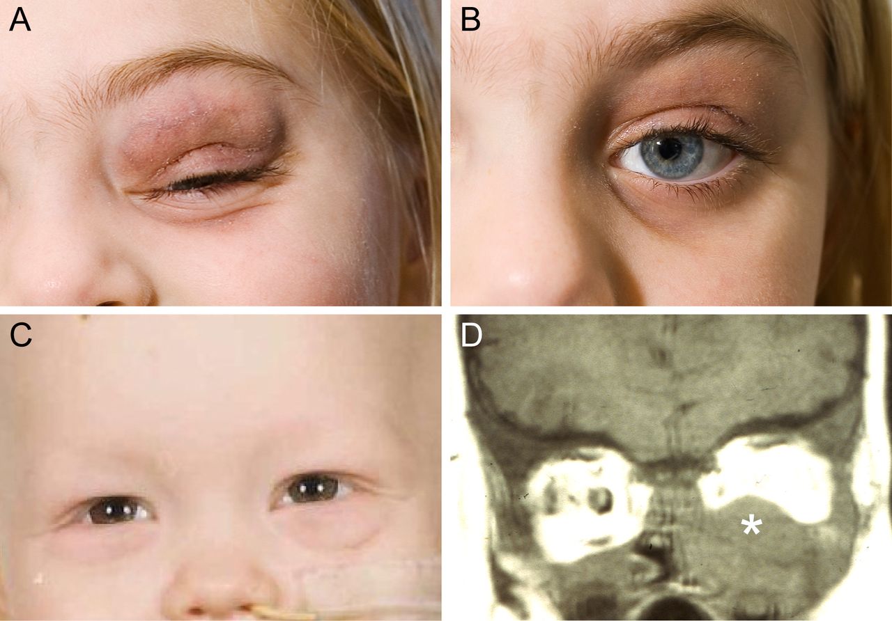
(A) 9-year-old girl (case 2) with left-sided proptosis, discolouration of the upper eye lid and ptosis. (B) Patient in (A) after 2 months of treatment. (C) 1-year-old boy (case 5) with left-sided proptosis and oedema of both eyelids. (D) MRI scan of the orbits (coronal view) of the patient in (C) shows a homogeneous mass involving the inferior half of the left orbit (asterisk).
Case 5 was a 1-year-old boy with fever and left-sided proptosis of a few weeks’ duration. He had oedema of both eyelids, bluish discolouration of the inferior lid and proptosis (4 mm) (figure 1C). CT and MRI showed a large homogeneous mass involving the inferior half of the left orbit, extending from the inferior lid to the orbital apex (figure 1D). Orbital and bone marrow biopsies revealed leukaemic infiltrates with neoplastic cells positive for Sudan black B, lysozyme, CD43 and CD45. The microscopic findings and the immunoprofile were consistent with AML, FAB type M5. Blood analysis showed pancytopenia. The patient had several blood transfusions and received chemotherapy (NOPHO AML 1993 protocol). He is still in remission, 18 years after diagnosis.
None of the three paediatric patients with primary orbital presentation of disease had any evidence of leukaemic infiltrates in the retina or choroid.
Clinical characteristics of secondary ophthalmic leukaemias
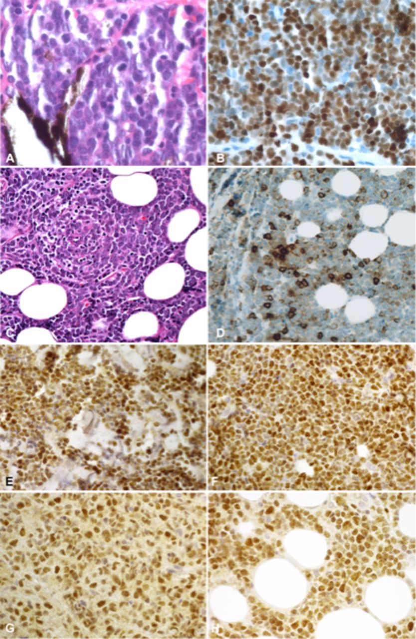
Six patients were diagnosed with systemic leukaemia before ophthalmic symptoms were present. Orbital/ocular manifestations were evident at a mean age of 49.2 years (range, 17–70 years). The leukaemic lesions were bilateral in one case, affected the left eye in three cases and the right eye in two. The average duration of symptoms before medical consultation was 5 weeks (range, 2–9 weeks). All patients with orbital tumours had proptosis, displacement of the eye and restricted eye mobility. Two patients with ocular manifestations had leukaemic infiltrates in the retina and subretinally; one of these patients also had involvement of the uvea and infiltration of the optic nerve. Two additional patients had infiltrates in the choroid, iris and anterior segment. Two patients with ocular lesions had pain from the eye; one also had blurred vision, and an examination revealed a visual acuity of 0.25, vitreous haze and papilloedema. One patient had a palpable mass. The lesions were diagnosed as BCP-ALL in two cases (figure 2A, B) and AML in three (figure 2C, D); one patient had a history of chronic lymphocytic leukaemia (CLL) with high-grade transformation to AML (table 1). All six patients received chemotherapy; two patients also received allogeneic bone marrow transplants, and two had radiotherapy. Four of the six patients died of relapse and/or disseminated disease 4–36 months after the onset of eye symptoms, one died of disseminated disease an unknown number of months after diagnosis and one died of heart failure 1.5 years after relapse.

(A) Lymphoblastic cells with irregular nuclei and sparse cytoplasm infiltrating the iris (H&E staining) in a patient with B-cell precursor acute lymphoblastic leukaemia (case 4); (B) lymphoblastic cells from case 4 are strongly immunoreactive for terminal deoxynucleotidyl transferase (TdT); (C) myeloblastic cells with an eosinophilic cytoplasm and indistinct cell boundaries and lymphocytes infiltrating the orbital fat tissue (H&E staining) in a patient with acute myeloid lekaemia Fab M1 (case 7); (D) myeloblastic cells from case 7 are strongly immunoreactive for myeloperoxidase; (E–H) expression of the MYB oncoprotein in ophthalmic leukaemic lesions from case 1, B-cell precursor acute lymphoblastic leukaemia (E), case 2, B-cell precursor acute lymphoblastic leukaemia (F), case 5, acute myeloid leukaemia Fab M5 (G), and case 7, acute myeloid leukaemia Fab M1 (H).
Expression of the MYB oncoprotein
Two of the three analysed BCP-ALLs showed strong nuclear immunostaining for the MYB oncoprotein in the majority of leukaemic cells (table 1 and figure 2E, F). Three of four analysed AML samples also had strong nuclear staining in the majority of neoplastic cells (table 1 and figure 2G, H). The MYB oncoprotein was not expressed in one BCP-ALL (case 3) and one AML (case 9).
Cytogenetic and molecular genetic characteristics
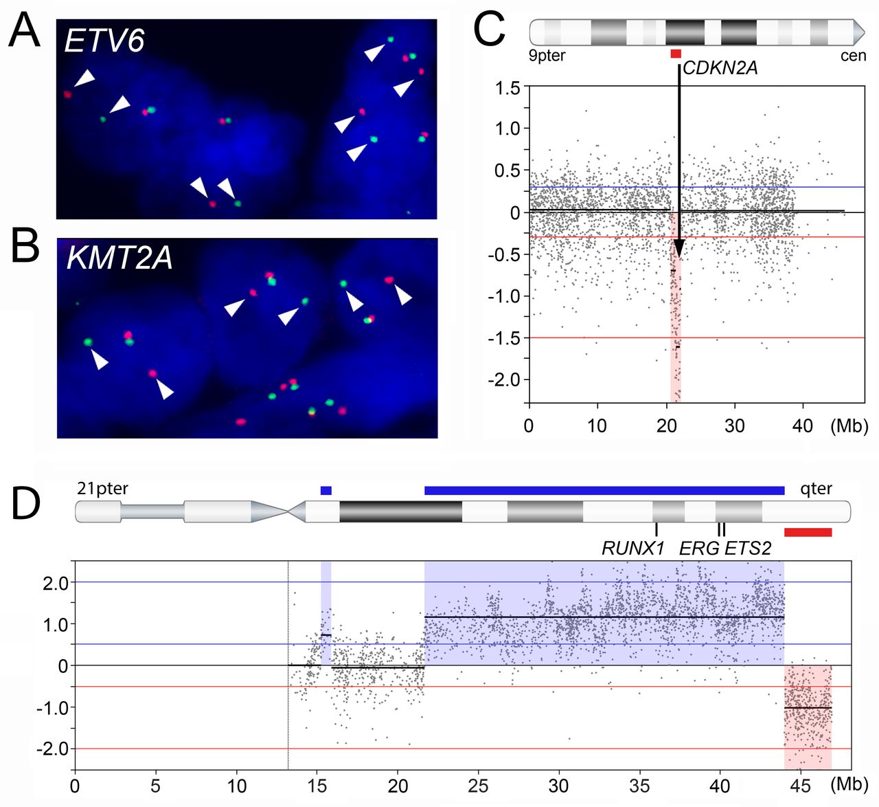
Cytogenetic information was available from three patients with BCP-ALL and four with AML. The karyotypic alterations are shown in table 1. Two of the three ALLs had abnormal karyotypes, and one had no cytogenetic changes (case 4). Case 2 had the classical t(12;21)(p13;q22) translocation associated with paediatric BCP-ALL, whereas case 1 had an uncommon t(2;3)(p11;q29) seen in a small subset of BCP-ALL. The case with the t(12;21) had a rearrangement of ETV6 consistent with an ETV6–RUNX1 gene fusion. FISH analysis also revealed an ETV6 rearrangement in case 3 (figure 3A); case 1 had no evidence of ETV6 rearrangement. Similarly, three of the four AMLs had abnormal karyotypes: case 5 had a t(9;11)(p22;q23) typical of the M5 subtype; case 6 had an inv(16)(p13q22), monosomy 7, and trisomy 11; and case 9 had a t(8;21)(q22;q22) resulting in a RUNX1–RUNX1T1 fusion. The fourth AML had an apparently normal karyotype (case 7). FISH analysis revealed that neither case 6 nor case 7 had any rearrangements of KMT2A, whereas case 5 had a rearranged KMT2A allele (figure 3B). Nucleotide sequence analysis revealed that case 8 (AML) had an FLT3 internal tandem duplication mutation and an exon 12 NPM1 mutation (data not shown).
FISH and arrayCGH analyses of acute leukaemias with ophthalmic manifestations. (A) FISH analysis showing a rearranged ETV6 allele (split red and green signals indicated by arrowheads) in a B-cell precursor acute lymphoblastic leukaemia (case 3). (B) FISH analysis showing a rearranged KMT2A allele (split red and green signals indicated by arrowheads) in a patient with acute myeloid leukaemia FAB M5 and a t(9;11) translocation (case 5). (C) ArrayCGH analysis showing homozygous loss of the tumour suppressor gene CDKN2A (arrow) in a B-cell precursor acute lymphoblastic leukaemia (case 3). (D) ArrayCGH analysis showing gain of 21q21.1–q22.3, including the RUNX1, ERG and ETS2 oncogenes, and loss of the terminal end of 21q in a B-cell precursor acute lymphoblastic leukaemia (case 2).
Genomic profiling
Genome-wide arrayCGH yielded analysable results from six of seven leukaemic patients with ophthalmic involvement (table 2), three of which had primary ophthalmic lesions (cases 1, 2 and 5). One BCP-ALL (case 1) and one AML (case 6) had no CNAs; the four other cases had an average of 3.3 CNAs per case (range 1–7) (table 2). One homozygous deletion, including the tumour suppressor CDKN2A, was detected in a BCP-ALL (case 3) (figure 3C). Case 2 (BCP-ALL) had gain of 21q21.1–q22.3, including the RUNX1, ERG and ETS2 oncogenes (figure 3D). Interestingly, this case had also gain of a 0.5 Mb segment in 12p13.2 and a breakpoint in ETV6, consistent with an ETV6–RUNX1 gene fusion. There were no high-level gene amplifications and no recurrent CNAs.
ArrayCGH analysis of seven cases of acute leukaemias with ophthalmic manifestations
Discussion
Here, we present a comprehensive clinical and genomic profiling study of nine leukaemias with orbital and/or ocular manifestations. Three were primary orbital manifestations of the leukaemia, representing all such cases histologically analysed in Denmark from 1980 to 2009. The remaining six cases had secondary orbital/ocular lesions. Four of our patients had BCP-ALL, and five had AML, one of which was originally a CLL. Transformation of CLL to high-grade AML is an uncommon event and is associated with a poor prognosis.9 Thus, all nine ophthalmic lesions in this study were acute leukaemias.
The average age at diagnosis of our patients with primary ophthalmic lesions was 5 years (24.2 years for all our patients), and there were similar numbers of males and females. The patients with orbital tumours presented with proptosis, displacement of the eye and reduced eye mobility. Two patients with ocular infiltrations had pain, and one also had reduced visual acuity. Notably, none of the paediatric patients with primary orbital manifestations had leukaemic infiltrates in the retina or choroid, whereas four of six patients with secondary ophthalmic leukaemias had such infiltrates. Taken together, our findings further emphasise the broad clinical spectrum of leukaemic lesions that may manifest in the eye or ocular region,1 3 4 6 7 10 11 and the need to consider leukaemia in the differential diagnosis of patients with proptosis, reduced mobility and/or displacement of the eye.1 4 6
The molecular mechanisms by which leukaemic cells give rise to extramedullary dissemination remain elusive. Thus, it is unclear why certain leukaemias present with orbital and/or ocular manifestations. There is, however, evidence suggesting that chemokines may be involved in organ-specific homing of neoplastic cells.12 Interestingly, ALL cells frequently express the chemokine receptors CXCR4 and CXCR3 and CLL cells the CXCR4 and CCR7 receptors.12 There is also a recent report demonstrating that α6 integrin, which frequently is overexpressed in ALL, interacts with laminin and mediates the migration of ALL cells to the central nervous system.13 Further studies are, however, needed to elucidate the exact mechanisms behind dissemination of acute leukaemias to the eye or ocular region.
The three patients with primary leukaemic orbital lesions were in complete remission 6, 9 and 18 years, respectively, after diagnosis. In previous studies, patients with orbital or ocular involvement of leukaemia had a poor prognosis and short overall survival since eye involvement often indicates recurrent disease.2 3 In our patients diagnosed with leukaemia before orbital/ocular lesions occurred, the median survival was 1.06 years (range, 2 months to 2 years). In a comprehensive study from the Children’s Oncology Group including 1459 paediatric patients with AML, those with involvement of orbital and central nervous system sites had a significantly better survival than patients with AML outside the central nervous system, those with leukaemia in the cerebrospinal fluid and those with no extramedullary leukaemia; the overall survival of the patients with AML with orbital involvement in this study was 92%.14
Cytogenetic data were available from three of four BCP-ALLs and four of five AMLs (table 1). Five cases had abnormal karyotypes and two had apparently normal karyotypes. The cytogenetic aberrations in these cases are similar to those in leukaemias without ophthalmic manifestations.15 16 FISH analysis revealed ETV6 rearrangements in two of three BCP-ALLs (table 1), consistent with ETV6-associated translocations. Indeed, case 2 had a t(12;21) translocation commonly associated with the ETV6–RUNX1 gene fusion seen in approximately 25% of paediatric ALLs.17 Patients with this fusion usually have a favourable prognosis.17 Similarly, FISH analysis revealed a rearrangement of KMT2A in one of three AML samples analysed. This case had a t(9;11)(p22;q23) known to result in a KMT2A–MLLT3 fusion.18 The prognostic significance of the t(9;11) is controversial. Recent studies suggest that this translocation carries an intermediate risk.19 20 Our patient with the t(9;11) was alive with no evidence of disease 18 years after diagnosis.
The genomic profiles of the orbital/ocular leukaemic lesions were further characterised by high-resolution arrayCGH (table 2). One BCP-ALL and one AML had no CNAs; the remaining four cases had rather quiet genomes. These findings are consistent with studies of leukaemic cells in bone marrow and/or peripheral blood,21–24 and with the observation that fusion gene-driven neoplasms often have few other genomic alterations.25 Notably, case 2 had gain of 21q21.1–q22.3, including the RUNX1, ERG and ETS2 oncogenes, and case 3 had a 1.5 Mb homozygous deletion in 9p21.3 including the CDKN2A tumour suppressor gene. This gene is frequently deleted in ALL and is associated with a poor prognosis and a poor response to treatment.26 27
MYB encodes a transcription factor that is important in the control of cell division, apoptosis and differentiation of haematopoietic stem/progenitor cells.28 29MYB is also an oncogene that is activated and overexpressed in subsets of acute leukaemias and in certain solid tumours.28–31 Here, we show for the first time that MYB is overexpressed in ophthalmic lesions in patients with acute leukaemias. In these cases, MYB is likely to be an important driver of leukaemogenesis and, therefore, also a potential therapeutic target. Further studies of MYB in acute leukaemias may therefore lead to better treatments for these malignancies.
In summary, we present the first comprehensive, integrated clinical, cytogenetic and genomic analyses of nine acute leukaemias with ophthalmic manifestations. These leukaemias did not differ significantly from those without clinically visible ophthalmic manifestations with regard to immunophenotype, cytogenetic aberrations, gene fusions and CNAs. Awareness of the clinical spectrum of leukaemic lesions of the eye or ocular region is important to quickly establish the correct diagnosis and commence prompt treatment.
Acknowledgments
We thank Therese Carlsson for excellent technical assistance.
References
Footnotes
Contributors LSS and SH planned and designed the study; LSS, A-CL and SC collected patient data; SH and SC performed pathology reviews; MP and MA performed experiments; LSS, MP, MA, SH, A-CL and GS collected and analysed data; LSS and GS drafted the manuscript. All authors contributed to the revision of the manuscript and approval of the final version.
Funding This study was supported by a grant from Synoptik-Fonden, Denmark and the Swedish Children’s Cancer Foundation.
Competing interests None declared.
Patient consent for publication Obtained.
Ethics approval This study was approved by the Local Scientific Ethics Committee (journal no. H-4-2013-003), the Danish Data Protection Agency (journal no. 2012-41-0747) and the Health Research Authority of England (REC Ref. 11/NW0759).
Provenance and peer review Not commissioned; externally peer reviewed.